BioCentral IBBioCentral IB
IB Biology · Theme A · A1.2

Nucleic acids

How life stores and copies information
Guiding questions

How does the structure of nucleic acids allow hereditary information to be stored?

How does the structure of DNA facilitate accurate replication?

Part one

Building blocks

A1.2.1 – A1.2.5
A1.2.1

The universal genetic material

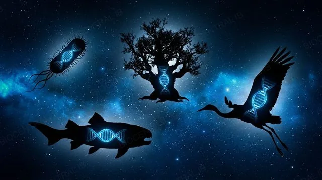
DNA (deoxyribonucleic acid) is the molecule that stores genetic information and passes it from one generation to the next.
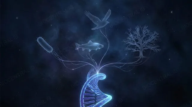
  • Every living organism — bacteria, archaea, fungi, plants, animals — uses DNA to store the instructions for building proteins and passing them on.
  • Nucleic acids have two jobs: storing genetic information, and carrying the code that tells the cell which proteins to build.
Why it's evidenceThis shared chemistry across every branch of life is one of the strongest pieces of evidence that all life descends from a common ancestor.
The exceptionSome viruses (influenza, SARS-CoV-2) carry RNA instead of DNA — this doesn't break the rule, because viruses aren't classified as living organisms: they have no cells and can't metabolise or reproduce on their own.
Silhouettes of a bacterium, tree, fish, and bird each containing a glowing DNA double helix
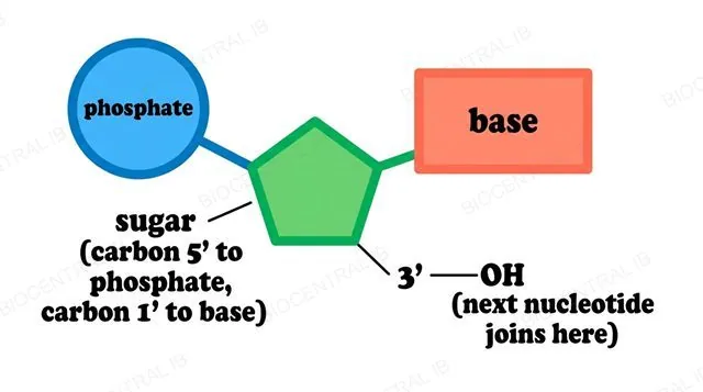
A labelled DNA nucleotide showing the phosphate, sugar, and base, with carbon positions marked
A1.2.2

The nucleotide

A nucleotide is the monomer of a nucleic acid — one phosphate group, one pentose (5-carbon) sugar, and one nitrogenous base.
  • The sugar's five carbons are numbered 1′ to 5′ — the prime symbol keeps them separate from the carbons inside the base.
  • Position matters: the base attaches to carbon 1′, the phosphate attaches to carbon 5′, and carbon 3′ carries a free —OH group — exactly where the next nucleotide in the chain will join.
IB conventionDiagrams draw the three parts as simple shapes: a circle for phosphate, a pentagon for the sugar, a rectangle for the base — the same notation used throughout this deck.
A1.2.3

The sugar-phosphate backbone

The backbone is the alternating chain of sugars and phosphates, joined by covalent bonds, running the length of a strand.
  • Nucleotides join by a condensation reaction — two molecules bond and a water molecule is released.
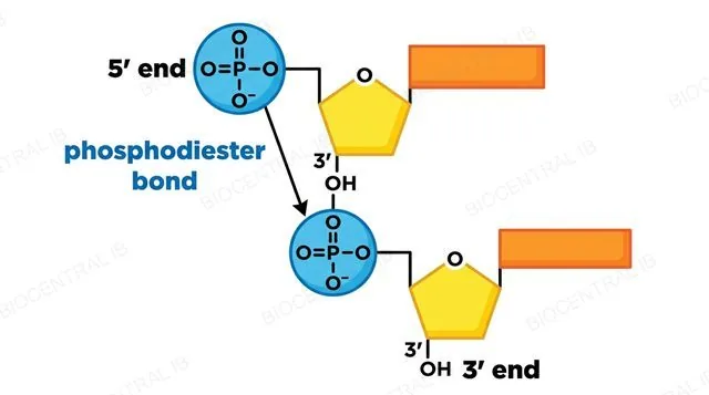
  • The new bond, a phosphodiester bond, forms between the —OH on carbon 3′ of the nucleotide already in the chain and the phosphate on carbon 5′ of the nucleotide being added.
Why it mattersBecause every nucleotide joins the same way, a finished strand has two different ends: a free phosphate at the 5′ end, and a free —OH at the 3′ end. The bases stick out sideways — the backbone itself never changes, so the information must be in the base order, not the backbone.
Two DNA nucleotides linked by a labelled phosphodiester bond, with the 5' and 3' ends marked
A long glowing DNA strand with a highlighted stretch representing a single gene
A1.2.4

The bases are the code

  • DNA has four bases:
    • Adenine (A)
    • Thymine (T)
    • Cytosine (C)
    • Guanine (G)
  • All four are nitrogen-containing rings that attach to carbon 1′ of the sugar the same way — which is why any base can sit at any position.
  • What differs is each base's shape and hydrogen-bonding pattern, which is why each has only one partner it can pair with.
A gene is a length of DNA whose base sequence carries the instructions for one specific product — usually a polypeptide — and so influences a characteristic of the organism.
Why it mattersBecause the order of bases is the information — exactly like letters in a word — a sequence read as ATGCAT means something different from TACGTA.
A single-stranded RNA polymer with ribose sugars and glowing bases
A1.2.5

RNA is built the same way

  • RNA is a polymer too, formed by condensation of RNA nucleotide monomers — the same reaction, releasing the same water, joined by the same phosphodiester bonds into a sugar-phosphate backbone.
Only two differencesThe sugar is ribose instead of deoxyribose, and uracil appears instead of thymine. The way the chain is put together is otherwise identical — and RNA is normally single-stranded, one chain instead of two.

Key vocabulary — building blocks

Worth being able to define in a single sentence each

Nucleotide
phosphate + sugar + base — the monomer of a nucleic acid.
Phosphodiester bond
the covalent bond joining one sugar's 3′ to the next nucleotide's 5′ phosphate.
Condensation reaction
a reaction that joins two molecules and releases water.
Backbone
the alternating chain of sugars and phosphates running the length of a strand.
Gene
a DNA sequence whose base order codes for one specific product.
5′ end / 3′ end
the two different ends of a strand — free phosphate vs free —OH.
Quick check

A student draws a nucleotide with the base attached to the sugar's 3′ carbon, and the phosphate attached to the 1′ carbon. What's wrong?

Nothing — that's correct
The base should attach to carbon 1′, and the phosphate to carbon 5′
Phosphates never attach directly to a sugar
The sugar should be drawn as a rectangle, not a pentagon
Correct answer: the positions are swapped. The base attaches to carbon 1′; the phosphate attaches to carbon 5′; carbon 3′ carries the free —OH where the next nucleotide joins.
Part two

The double helix and its jobs

A1.2.6 – A1.2.10
A1.2.6

The double helix

  • DNA is two strands wound around each other.
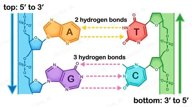
  • Complementary base pairing fixes A with T and C with G — the shapes and hydrogen-bonding atoms of the bases allow no other combination.
  • Hydrogen bonds hold the pairs together: 2 bonds between A and T, 3 bonds between C and G — weak alone, but millions together hold the molecule firmly while still allowing it to be "unzipped" by enzymes.
AntiparallelThe two strands run in opposite directions — where one has its 5′ end, the other has its 3′ end. Only in this arrangement do the bases line up facing each other so hydrogen bonds can form.
A-T base pair with two hydrogen bonds and G-C base pair with three hydrogen bonds, strands shown running in opposite directions
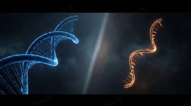
Side-by-side comparison of a thick DNA double helix and a thin single RNA strand
A1.2.7

DNA compared with RNA

DNA
  • Sugar: deoxyribose
  • Two strands (double helix)
  • Very long, stable, long-lived
  • Kept in the nucleus (also mitochondria/chloroplasts)
RNA
  • Sugar: ribose; base U not T
  • One strand
  • Shorter, often broken down after use
  • Made in the nucleus, works in the cytoplasm
A useful way to hold itDNA is the master copy kept in the archive; RNA is the photocopy you take to the workshop.
A1.2.8

Job one: replication

  • Replication (copying): hydrogen bonds break and the two strands separate; each old strand acts as a template, so free nucleotides pair up according to the rule.
  • Because each base has only one possible partner, the new strand can only be an exact complement — producing two identical DNA molecules, each made of one old strand and one new one.
Why it mattersThis “semi-conservative” copying — each new molecule keeps one original strand — is how genetic information passes accurately from a cell to its daughter cells.
A DNA double helix unzipping with new complementary strands forming along each template strand
An mRNA strand being synthesized along a DNA template strand, with RNA polymerase shown building the new strand
A1.2.8

Job two: transcription

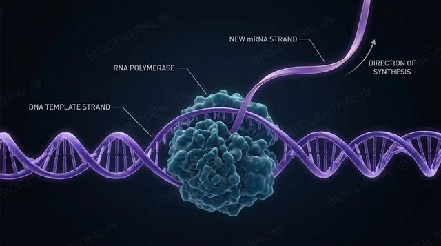
  • Transcription (expression): one DNA strand acts as a template for a new mRNA strand, built by the same base-pairing rule — except A on the DNA pairs with U on the RNA, since RNA has no thymine.
  • Unlike replication, only a short stretch of DNA — one gene — is transcribed at a time, and only one of the two strands is used as the template.
Why it mattersThe finished mRNA carries a faithful copy of the gene’s message out to the ribosome, where it directs protein synthesis — this is how the code stored in DNA gets expressed as a working protein.
An impossibly long strand of DNA coiling into the distance toward a starfield
A1.2.9

Vast storage

  • Any of the four bases can sit at any position — no chemical rule restricts the order.
  • A strand n bases long has 4ⁿ possible sequences, and the number explodes fast: 10 bases gives about a million sequences; a modest 1,000-base gene gives 4¹⁰⁰⁰, a number far larger than the atoms in the observable universe.
Why it mattersA human genome — about 3 billion base pairs — has vastly more possible sequences than there have ever been organisms, which is why every species, and every individual, can have its own unique sequence.
A1.2.10

One shared genetic code

The genetic code is the set of rules by which a sequence of bases is read in triplets and translated into amino acids.
  • The code is universal — the same triplet stands for the same amino acid in bacteria, a mushroom, an oak tree, and you.
  • There's nothing chemically necessary about these assignments — no reason the triplet GGC has to mean glycine.
  • If life began independently more than once, we'd expect different codes.
A practical consequenceBecause the code is shared, a human gene transferred into a bacterium is read using the same rules — the basis of genetic engineering (human insulin made by bacteria). The code is almost universal: a few minor variations exist (e.g. mitochondria), but the code is overwhelmingly conserved.
A single glowing DNA helix branching outward into silhouettes of different life forms
Higher Level

Extension

A1.2.11 – A1.2.15 · not on the SL course
A1.2.11 · HL

Directionality: 5′ and 3′

  • A strand is not symmetrical.
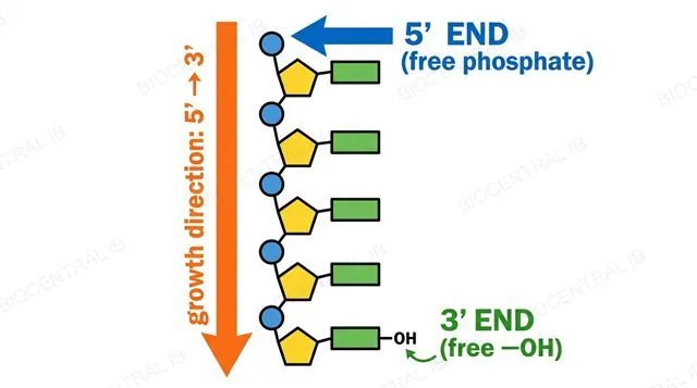
  • New nucleotides can only be added to the 3′ end, because the new phosphodiester bond forms with that free —OH — so a strand is always built in the 5′ → 3′ direction, exactly as shown opposite.
In replicationDNA polymerase only works 5′ → 3′. Because the two templates are antiparallel, only one new strand — the leading strand — is made continuously toward the replication fork; the other, the lagging strand, is made in short pieces.
In transcription & translationRNA polymerase also works 5′ → 3′, reading the DNA template 3′ → 5′. The ribosome then moves along the finished mRNA from its 5′ end toward its 3′ end, reading codon by codon.
A DNA strand with the 5' phosphate end and 3' hydroxyl end explicitly labelled, with an arrow showing the 5' to 3' growth direction
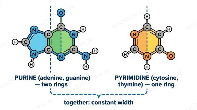
A larger two-ring purine molecule beside a smaller single-ring pyrimidine molecule, both labelled
A1.2.12 · HL

Purines, pyrimidines, and helix stability

  • The four bases fall into two structural groups:
    • Purines (adenine, guanine) — two fused rings, larger
    • Pyrimidines (cytosine, thymine, and uracil in RNA) — single ring, smaller
  • In DNA, a purine always bonds to a pyrimidine.
Why it mattersEvery base pair is the same width, so the two backbones stay a constant distance apart along the whole molecule — the helix has a uniform diameter that doesn't bulge or pinch, which keeps the hydrogen-bonding atoms exactly the right distance apart to bond.
A1.2.13 · HL

The nucleosome

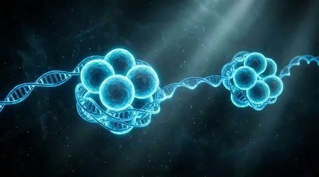
A nucleosome is a DNA molecule wound about twice around a core of eight histone proteins, held by an additional H1 histone.
  • A human cell holds about 2 metres of DNA inside a nucleus a few micrometres across.
  • During interphase, DNA exists as chromatin — DNA plus histones, arranged as a repeating "beads on a string" of nucleosomes, connected by stretches of linker DNA.
How it's held onThe backbone is negatively charged (phosphates); histones are rich in positively charged amino acids. The ionic attraction doesn't depend on base sequence, so histones can bind anywhere — packaging the DNA while also affecting whether a gene can be transcribed.
A DNA strand wound around a histone octamer, forming a beads-on-a-string nucleosome structure
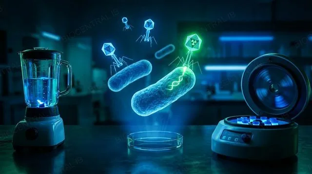
A laboratory blender and centrifuge with glowing bacteriophage particles infecting bacteria
A1.2.14 · HL

The Hershey-Chase experiment

  • The question: a bacteriophage is only protein (coat) and DNA (inside) — which one is the genetic material it injects into a bacterium?
  • The tool: the phage's protein contains sulfur but no phosphorus; DNA contains phosphorus but no sulfur — so radioactive ³⁵S labelled protein, and ³²P labelled DNA.
  • After infecting bacteria, the mixture was blended to shear off empty coats, then centrifuged: heavy bacteria formed a pellet, light coats stayed in the supernatant.
Result³⁵S was found mainly in the supernatant (protein stayed outside); ³²P was found mainly in the pellet (DNA entered the cells) — strong evidence DNA is the genetic material. The separation wasn't perfect, but the clear difference between batches made the case — good science reports the untidiness too.
A1.2.15 · HL

Chargaff's data

  • In the 1940s-50s, Erwin Chargaff measured base percentages across many species.
  • Within any one species, A ≈ T and G ≈ C — a purine-to-pyrimidine ratio of about 1:1.
  • But the proportions weren't the same between species — humans have far more A and T than G and C, while E. coli has roughly equal amounts of all four.
FalsifiabilityThe prevailing tetranucleotide hypothesis predicted every organism's DNA should be a monotonous 25%-each repeat — a testable, falsifiable claim. Chargaff's data falsified it: DNA varied between species, so it was complex enough to carry information after all. The 1:1 ratio also hinted that bases are paired — exactly what Watson and Crick used to arrive at A-T and C-G pairing.
Human A≈31% T≈29% · E. coli A≈25% T≈24%
A laboratory still-life with glowing colored spheres balanced in pairs, representing base composition data
Quick check · HL

DNA polymerase can only add a new nucleotide to a strand's free 3′ —OH. In which direction is a new strand always built?

3′ → 5′
5′ → 3′
It depends on which strand is being copied
Both directions at once
Correct answer: 5′ → 3′, always. Because the new phosphodiester bond forms with the free —OH on carbon 3′, growth can only happen at that end — which is also why the lagging strand has to be built in short, separate pieces during replication.

Key vocabulary — HL extension

Worth being able to define in a single sentence each

Purine
a base with two fused rings: adenine or guanine.
Pyrimidine
a base with a single ring: cytosine, thymine, or uracil.
Chromatin
DNA together with its histones, as it exists during interphase.
Histone
a positively charged protein that DNA winds around.
Nucleosome
DNA wound around a histone octamer, held by an H1 histone.
Radioisotope
a detectable form of an element that behaves normally in reactions.

Where this shows up again

D1.1 · DNA replication
How is the base sequence copied when DNA replicates before cell division?
D1.2 · Protein synthesis
How is the information in a DNA base sequence used to build a specific protein?
D1.3 · Mutation
How do mutations change the base sequence, and what can be the consequences?
A2.3 · Viruses
How do some viruses use RNA instead of DNA as their genetic material?

A1.2 Nucleic acids — one-page recap

Screenshot this slide to revise from

The nucleotide
  • Phosphate + sugar + base
  • Base on 1′, phosphate on 5′, —OH on 3′
Backbone & code
  • Phosphodiester bonds via condensation
  • Base order is the information
DNA vs RNA
  • Deoxyribose/ribose; T vs U
  • Double- vs single-stranded
The double helix
  • A-T (2 H-bonds), C-G (3 H-bonds)
  • Antiparallel strands
Replication & code
  • Each strand templates its complement
  • Genetic code ≈ universal → common ancestry
HL · Structure & evidence
  • Strands build 5′→3′ only
  • Purine+pyrimidine = constant width
  • Nucleosome: DNA + 8 histones

Four letters. Vast information.

That's the whole code of life.
A1.2 Nucleic acids · BioCentral IB
Use ↓ ↑ or click to navigate
01 / 20