BioCentral IBBioCentral IB
IB Biology · Theme A · A2.2

Cell Structure

The unit of life — and how we see inside it
Guiding questions

What are the features common to all cells and the features that differ?

How is microscopy used to investigate cell structure?

Part one

The cell as the unit of life

A2.2.1 · A2.2.4
A2.2.1

Every organism is built from cells

A cell is the smallest unit that is both structurally complete (a membrane enclosing genetic material) and functionally complete (able to carry out every process of life).
  • Every organism — from a single bacterium to the roughly 30 trillion cells of a human body — is built from cells.
  • This is biology's basic organising principle: to understand any tissue, organ, or organism, you ultimately have to understand the cells that build it.
Why it mattersBecause cell theory sets both requirements, a structure lacking either a membrane or genetic material cannot be classified as a cell on its own.
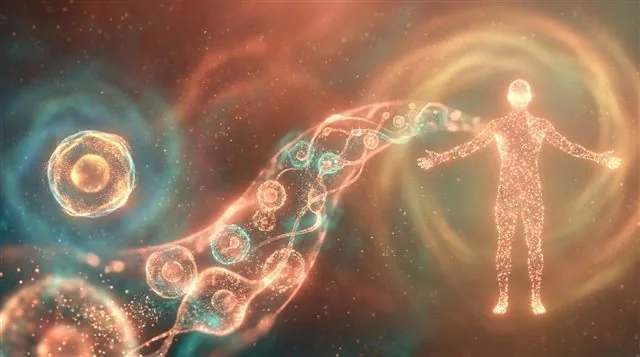
Artistic scale progression from a single cell to a full multicellular body
A2.2.4

Four structures every cell shares

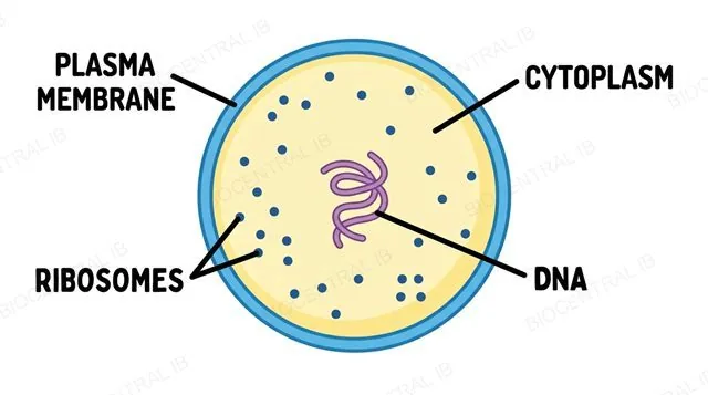
Despite their differences, every cell — prokaryotic or eukaryotic — shares four structures: a plasma membrane, cytoplasm, ribosomes, and DNA.
  • Every cell shares the same universal toolkit:
    • Plasma membrane — controls what enters and leaves the cell
    • Cytoplasm — the jelly-like material inside where reactions occur
    • Ribosomes — build proteins by translating the genetic code
    • DNA — carries the instructions the cell needs
  • Everything else you will meet — a nucleus, a cell wall, membrane-bound organelles — is a variation added on top of this shared, universal toolkit.
Why it mattersThese four features are the minimum a structure needs before biologists will call it a living cell at all.
Generalised cell labelled with plasma membrane, cytoplasm, ribosomes, and DNA
Part two

Seeing the invisible: microscopy

A2.2.2 · A2.2.3
A2.2.2

Calculating magnification

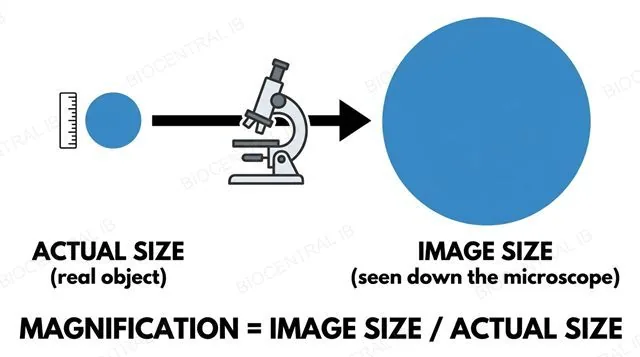
Magnification is how many times larger an image is than the real, actual object — calculated as image size ÷ actual size.
  • A scale bar labelled 5 μm that measures 25 mm on a printed micrograph gives a magnification of ×5000, once both measurements are converted to the same unit (25 mm = 25,000 μm; 25,000 ÷ 5 = 5000).
  • The same equation rearranges to find a structure's real size once the magnification is known: actual size = image size ÷ magnification.
Why it mattersThe single most common error is mixing units — always convert millimetres and micrometres before dividing (1 mm = 1000 μm = 1,000,000 nm).
Diagram illustrating the magnification formula: image size divided by actual size
A2.2.3

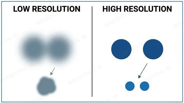
Resolution is the real limit

Resolution is the smallest distance between two points at which they can still be seen as separate, rather than blurring into one.
  • Magnification and resolution are often confused, but only resolution sets the real limit on detail — an image can be magnified as much as you like, but if the detail was never resolved to begin with, magnifying it further just produces a bigger blur.
  • A light microscope resolves to about 200 nm and can view living, coloured cells; an electron microscope resolves to about 0.1–2 nm, revealing ultrastructure — membranes, ribosomes, cristae — but only in fixed, dehydrated, naturally black-and-white samples viewed in a vacuum.
Why it mattersResolution, not magnification, is what actually determines how much detail any instrument can reveal.
Two panels comparing low resolution (blurred blob) versus high resolution (two separate points)
A2.2.3 continued

Freeze fracture: splitting the membrane

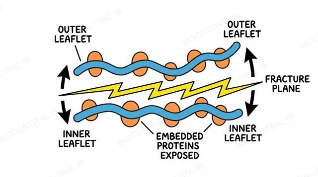
Freeze fracture freezes a specimen solid and then physically cracks it, splitting membranes along their weak internal plane.
  • Membranes tend to fracture down the middle, between their two phospholipid layers, rather than across them — exposing the proteins embedded within the membrane itself.
  • The exposed surfaces are coated with a thin layer of metal and imaged, revealing the size, shape and distribution of embedded proteins that no stain could show directly.
Why it mattersFreeze fracture is the technique that first showed biologists membrane proteins actually exist, embedded within the lipid bilayer rather than sitting on its surface.
Labelled diagram of freeze fracture: a membrane split along its fracture plane, exposing embedded proteins in the outer and inner leaflets
A2.2.3 continued

Cryo-EM: freezing molecules mid-shape

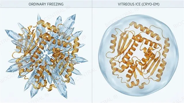
Cryogenic electron microscopy (cryo-EM) freezes a specimen so fast that ice crystals have no time to form.
  • Ordinary freezing forms ice crystals that distort or destroy delicate structures; cryo-EM's extremely rapid freezing traps water as amorphous, glass-like ice instead, leaving molecules close to their natural shape.
  • Because the specimen never needs staining, dehydrating or chemical fixation, cryo-EM can resolve the fine three-dimensional structure of large protein complexes and viruses at near-atomic detail.
Why it mattersCryo-EM's main advantage is preserving a molecule's natural shape — most other electron microscopy techniques require preparation steps that can distort delicate structures.
Diagram illustrating cryo-EM: a specimen flash-frozen in vitreous ice, preserving its natural molecular shape, compared with ice-crystal damage from ordinary freezing
A2.2.3 continued

Immunofluorescence: lighting up one protein

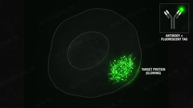
Immunofluorescence attaches a fluorescent tag to an antibody that binds only to one specific target protein.
  • The tagged antibody is added to a light-microscope sample, where it seeks out and binds only its matching protein — every other structure in the cell stays dark.
  • Viewed under fluorescent light, the tag glows exactly where that one protein is located, revealing its precise position inside or on the cell.
Why it mattersImmunofluorescence's main advantage is specificity: unlike a general stain, it identifies the location of one particular protein among thousands of others in the same cell.
Diagram of immunofluorescence: a fluorescent-tagged antibody binding a specific target protein inside a cell, glowing under fluorescent light while the rest of the cell stays dark
Part three

Prokaryotes, eukaryotes, and single-celled life

A2.2.5 · A2.2.6 · A2.2.7
A2.2.5

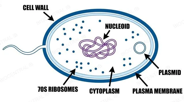
Prokaryote cell structure

A prokaryotic cell (bacteria and archaea) has no nucleus and no membrane-bound organelles.
  • Its DNA is a single circular molecule lying free in a region called the nucleoid, and its ribosomes are 70S — smaller than a eukaryotic cell's.
  • A rigid cell wall (peptidoglycan, in the Gram-positive bacteria this course requires, such as Bacillus and Staphylococcus) sits outside the plasma membrane for shape and protection.
  • Many prokaryotes also carry small circular plasmids of extra DNA, and some have a whip-like flagellum for movement.
Why it mattersProkaryote structure varies between species, but you are not required to know the details of every variation.
Labelled prokaryotic cell showing cell wall, plasma membrane, nucleoid, ribosomes, plasmid, and flagellum
A2.2.6

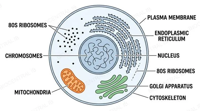
Eukaryote cell structure

A eukaryotic cell (animals, plants, fungi, protists) has a nucleus and membrane-bound organelles.
  • Its DNA is linear, associated with histone proteins, and enclosed within the nucleus's own double membrane.
  • Membrane-bound organelles — the mitochondrion, endoplasmic reticulum, Golgi apparatus — each provide their own internal environment suited to a specific job, an arrangement called compartmentalisation.
  • Its ribosomes are 80S, larger than a prokaryote's 70S.
Why it mattersCompartmentalisation lets incompatible reactions run simultaneously in the same cell, each isolated in its own organelle.
Labelled eukaryotic animal cell showing nucleus, mitochondria, ER, Golgi apparatus, and cytoskeleton
A2.2.7

One cell doing everything

A unicellular organism — such as Amoeba, Paramecium, Chlamydomonas, or a yeast cell — is a single cell that carries out every process of life by itself.
  • Homeostasis, metabolism, nutrition, movement, excretion, growth, response to stimuli, and reproduction — nothing is delegated to a neighbouring cell, because there is no neighbouring cell.
  • Every structure the organism has must serve one or more of these functions directly.
Why it mattersThis is the sharpest possible contrast with a multicellular organism, where individual cells specialise and share the workload.
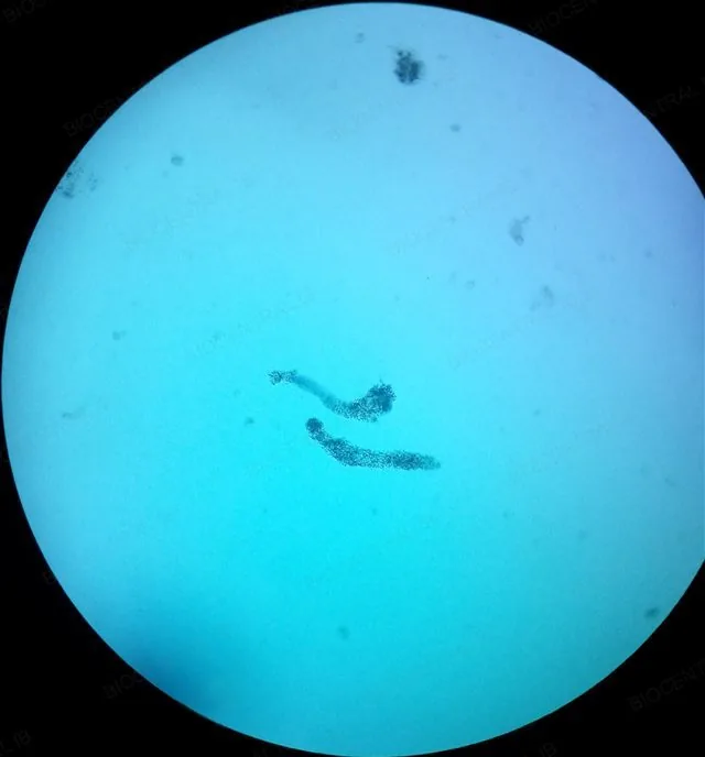
Amoeba proteus viewed under a light microscope, showing pseudopodia extending from the cell surface
Fritzmann2002 / Wikimedia, CC BY-SA 4.0
Amoeba proteus (light microscopy)
Quick check

A student measures a scale bar as 20 mm long on a micrograph. The scale bar is labelled 2 μm. What is the magnification?

×10
×10,000
×40
×2,000
Correct answer: ×10,000. Convert to the same unit first (20 mm = 20,000 μm), then divide: 20,000 ÷ 2 = 10,000. Skipping the unit conversion — dividing 20 by 2 — is the most common version of this mistake.
Part four

Diversity, exceptions, and reading cells

A2.2.8 · A2.2.9 · A2.2.10 · A2.2.11
A2.2.8

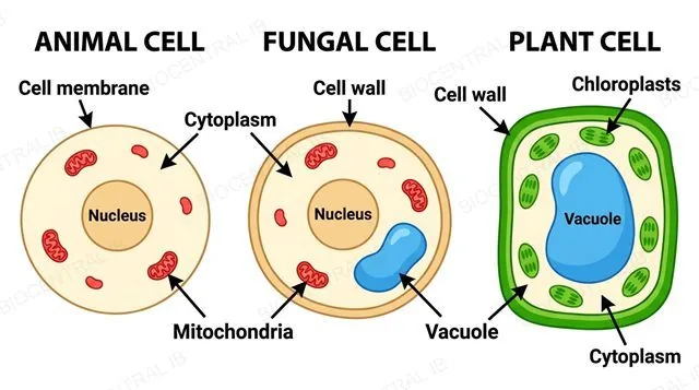
Animal, fungal, and plant cells

Animal, fungal, and plant cells all share the eukaryotic core — yet differ in several structural details layered on top of it.
  • Cell wall differs by kingdom:
    • Animal — no cell wall
    • Fungal — chitin wall
    • Plant — cellulose wall
  • Only plant cells carry chloroplasts and a large, permanent central vacuole.
  • Centrioles are typical of animal cells but absent from most fungal and plant cells.
  • Nutrition also differs by kingdom:
    • Animals — ingest food
    • Fungi — absorb it after external digestion
    • Plants — photosynthesise
Why it mattersThese are differences layered on top of the shared eukaryotic plan from A2.2.6, not a different plan altogether.
Animal, fungal, and plant cells compared side by side
A2.2.9

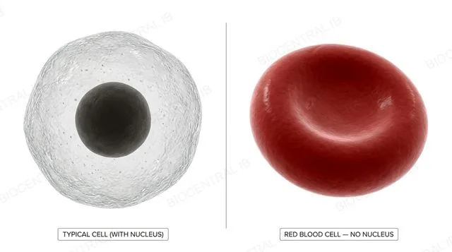
Red blood cells lose their nucleus

A mature mammalian red blood cell has no nucleus — one clear example of atypical cell structure defined by the number of nuclei a cell contains.
  • As a red blood cell matures in the bone marrow, it expels its nucleus along with most other organelles, becoming a flattened, biconcave disc almost entirely filled with haemoglobin.
  • The extra internal space raises the amount of haemoglobin — and so the amount of oxygen — each cell can carry, while the biconcave shape increases surface area for gas exchange.
Why it mattersLosing its nucleus means a mature red blood cell can no longer divide or make new proteins — it survives roughly 120 days before the body replaces it.
Cross-section of a mature red blood cell, biconcave disc shape, showing no nucleus and packed with haemoglobin, next to a typical nucleated cell for comparison
A2.2.9 continued

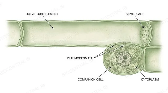
Sieve tubes lose their nucleus but stay alive

A phloem sieve-tube element also loses its nucleus at maturity — but unlike a red blood cell, it survives only because a neighbouring cell keeps it alive.
  • Losing its nucleus and most organelles clears the tube's interior, letting sugars (mainly sucrose) flow through pores in its end walls, called sieve plates, with minimal obstruction.
  • An adjacent, fully nucleate companion cell stays connected to the sieve-tube element through plasmodesmata, supplying it with ATP and proteins it can no longer make for itself.
Why it mattersA sieve-tube element cannot survive alone — its atypical structure only works because of this permanent partnership with its companion cell.
Labelled diagram of a phloem sieve-tube element with no nucleus, connected via plasmodesmata to an adjacent nucleate companion cell, with sieve plates at each end
A2.2.9 continued

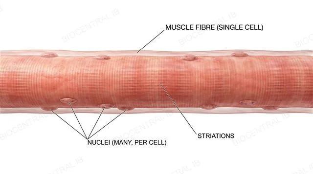
One giant cell, many nuclei

A skeletal muscle fibre is a single, unusually large cell containing many nuclei, formed by the fusion of many separate embryonic cells into one.
  • During development, many individual myoblasts fuse together into one continuous fibre — and each keeps its own nucleus rather than the fibre reducing to just one.
  • A fibre can run centimetres long and carry hundreds of nuclei spread along its length, each supporting protein synthesis in the section of cytoplasm nearest to it.
Why it mattersA muscle fibre needs far more protein-making capacity than a single nucleus could support — multiple nuclei let it sustain its huge volume of contractile protein.
Labelled diagram of a long skeletal muscle fibre formed by fusion of myoblasts, showing many nuclei spread along its length beneath the cell membrane
A2.2.9 continued

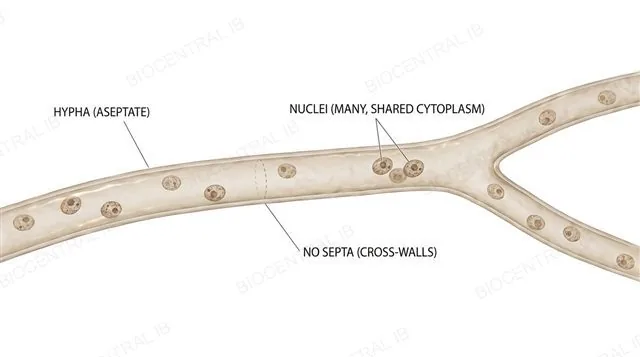
No walls between nuclei

Aseptate fungal hyphae are long tubes of continuous cytoplasm containing many nuclei, with no cross-walls dividing one region from the next.
  • Most fungal hyphae are divided into compartments by cross-walls called septa; aseptate hyphae lack these, so nuclei and cytoplasm move freely along the whole length of the hypha.
  • This uninterrupted structure lets resources and signalling molecules travel rapidly through the fungus, supporting fast growth at the hyphal tip.
Why it mattersAseptate hyphae, like the red blood cell, phloem sieve-tube element and muscle fibre, show that nucleus number is set by function, not by a fixed one-nucleus-per-cell rule.
Labelled diagram of an aseptate fungal hypha, a continuous tube of cytoplasm with many nuclei scattered along its length and no septa dividing it
A2.2.10

Reading a micrograph

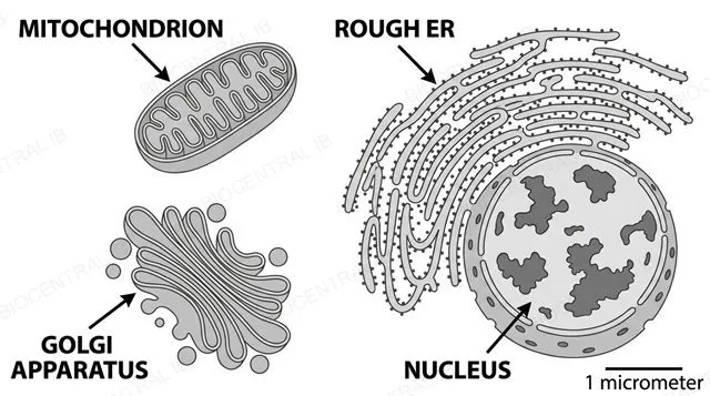
You are expected to identify cells and organelles directly from real light and electron micrographs.
  • Visual identification cues:
    • Double membrane with internal folds → mitochondrion or chloroplast
    • Stack of flattened, unconnected membrane sacs → Golgi apparatus
    • Membrane studded with small dark dots → rough ER
    • Large, round structure with darker patches → nucleus
  • Scale is a clue, not just a calculation — a structure a few nanometres across cannot be a nucleus, and a structure several micrometres across cannot be a ribosome.
Why it mattersUse the numbers alongside the shape: size rules out wrong answers as fast as appearance does.
Labelled electron-micrograph-style diagram identifying mitochondrion, Golgi apparatus, rough ER, and nucleus
A2.2.11 · Practical skill

Drawing what you actually see

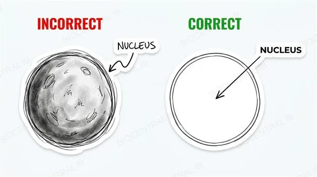
A scientific drawing records only what is actually visible in one specific micrograph — nothing added from memory.
  • Draw only what you can see, using single, clear, unbroken lines with no shading or sketchy repeated strokes.
  • Keep proportions accurate — if one organelle is twice the size of another in the micrograph, it should be twice the size in your drawing.
  • Label lines must be thin, straight, and ruler-drawn, touching the structure they label and never crossing another line.
  • Always include a title and the magnification or a scale bar.
Why it mattersExaminers mark proportion, line quality, and correct, function-linked labelling — not artistic detail.
Comparison of an incorrect shaded sketch versus a correct single-line labelled drawing of an organelle

Key vocabulary — cell structure

Worth being able to define in a single sentence each

Magnification
How many times larger an image is than the real, actual object.
Resolution
The smallest distance between two points at which they can still be seen as separate.
Prokaryotic cell
A cell with no nucleus and no membrane-bound organelles; DNA lies free in the nucleoid.
Eukaryotic cell
A cell with a membrane-bound nucleus and membrane-bound organelles.
Nucleoid
The region of a prokaryotic cell where the circular DNA is located; not membrane-bound.
Compartmentalisation
The division of a eukaryotic cell into separate membrane-bound regions, each with its own environment.
Part five · HL only

Origins and specialisation

A2.2.12 · A2.2.13 · A2.2.14
A2.2.12 · HL

Mitochondria were once free-living bacteria

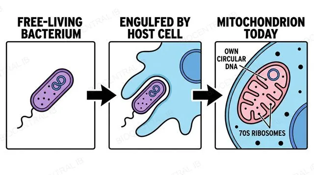
Mitochondria — and, in plants and algae, chloroplasts — were once free-living prokaryotes engulfed by a host cell, surviving as a permanent endosymbiosis.
  • The evidence lies in features unnecessary for an organelle but expected of a bacterium:
    • A double membrane (the inner one is the engulfed bacterium's own)
    • Their own small circular DNA, separate from the nucleus
    • 70S ribosomes, matching a bacterium's rather than the cell's 80S
    • The ability to grow and divide by binary fission independently of the rest of the cell
Why it mattersA wide range of independent observations converging on one explanation is the hallmark of a strong scientific theory.
Three-step diagram of endosymbiotic theory: free-living bacterium, engulfment by host cell, mitochondrion today
A2.2.13 · HL

Same genome, different jobs

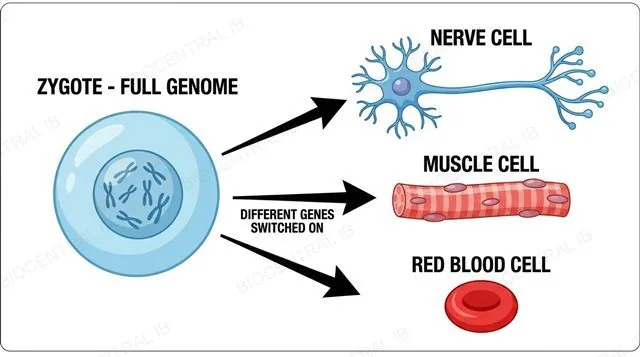
Cell differentiation is the process by which a cell becomes structurally and functionally specialised by expressing only a subset of its genome.
  • Every cell in a multicellular organism carries a complete copy of the same genome, inherited from the same zygote — yet a nerve cell looks nothing like a muscle cell or a red blood cell.
  • The difference is not which genes a cell has but which genes are switched on: a muscle cell expresses contractile-protein genes; a nerve cell expresses ion-channel genes and grows a long extension; a red blood cell expresses haemoglobin genes heavily, then discards its own nucleus.
Why it mattersDifferentiation is what makes a division of labour between cells possible — the multicellular counterpart of one cell doing everything.
Diagram showing a zygote branching into a nerve cell, muscle cell, and red blood cell via different gene expression
A2.2.14 · HL

Multicellularity evolved again and again

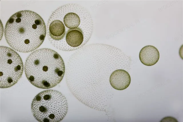
Multicellularity — an organism built from many cooperating cells — evolved independently multiple times: separately in animals, plants, fungi, and several algal groups.
  • This repeated, independent origin is convergent evolution.
  • Cells that stay attached after dividing, rather than separating, can specialise for different jobs — feeding, protection, reproduction — instead of every cell doing everything itself, a division of labour that can make the whole organism more efficient than the sum of independent single cells.
Why it mattersA larger multicellular body can access resources or avoid predation a single cell cannot — but only once differentiation lets its cells take on coordinated roles.
Volvox, a real colonial green alga made of thousands of cooperating cells
Frank Fox / mikro-foto.de, CC BY-SA 3.0

Key vocabulary — origins & specialisation

HL only — worth being able to define in a single sentence each

EndosymbiosisHL
A permanent, mutually beneficial relationship in which one organism lives inside the cell of another.
Cell differentiationHL
A cell becoming structurally and functionally specialised by expressing a specific subset of its genome.
MulticellularityHL
An organism built from many cells that cooperate and divide labour between them.
Convergent evolutionHL
The independent evolution of similar features in separate lineages, such as multicellularity arising repeatedly.
Quick check · HL

Which of these is NOT evidence used to support the endosymbiotic theory of mitochondria?

Mitochondria have their own small circular DNA
Mitochondria have a double membrane
Mitochondria are the same size in every cell type
Mitochondria can divide by binary fission independently of the cell
Correct answer: mitochondrial size. The theory's evidence is the double membrane, own circular DNA, 70S ribosomes, and independent binary fission — features unnecessary for an organelle but expected of a free-living bacterium. Size is not part of that evidence.

Where this shows up again

B2.1 · Membranes and membrane transport
How does the structure of the plasma membrane control the exchange of substances that a cell's surface area allows?
B2.3 · Cell specialization
How do genetically identical cells become specialised into different cell types in a multicellular organism?
C1.2 · Cell respiration
How do mitochondria use their internal structure to release energy in aerobic cell respiration?
A2.3 · Viruses
How do viruses differ from cells, given that they lack cytoplasm and cannot carry out the functions of life alone?

A2.2 Cell Structure — one-page recap

Screenshot this slide to revise from

Seeing cells
  • Magnification: image size ÷ actual size.
  • Resolution — not magnification — sets the detail limit (LM ~200 nm; EM ~0.1–2 nm).
Universal toolkit
  • Every cell: plasma membrane, cytoplasm, ribosomes, DNA.
  • Everything else is added on top of this shared plan.
Prokaryote vs eukaryote
  • Prokaryote: no nucleus, 70S ribosomes, nucleoid DNA.
  • Eukaryote: nucleus + organelles, 80S ribosomes.
Diversity & exceptions
  • Animal/fungal/plant cells differ in wall, vacuole, chloroplasts.
  • RBCs, muscle fibres, hyphae all break the generalised model.
HL · Origins
  • Mitochondria evolved by endosymbiosis.
  • Evidence: double membrane, own DNA, 70S ribosomes, binary fission.
HL · Specialisation
  • Differentiation: same genome, different genes switched on.
  • Multicellularity evolved independently, many times over.

One genome. A thousand different jobs.

Next: how the plasma membrane you just met actually controls what crosses it.
A2.2 Cell Structure · BioCentral IB
Use ↓ ↑ or click to navigate
01 / 20