BioCentral IBBioCentral IB
IB Biology · Theme B · B1.2

Proteins

How a string of twenty amino acids folds into enzymes, haemoglobin and silk — and why changing a single link can reshape the whole machine.
Guiding questions

What is the relationship between amino acid sequence and the diversity in form and function of proteins?

How are protein molecules affected by their chemical and physical environments?

Part one

Amino acids and the polypeptide chain

B1.2.1 – B1.2.5
B1.2.1

Twenty amino acids, one shared backbone

An amino acid is a molecule with a central (alpha) carbon bonded to an amino group, a carboxyl group, a hydrogen atom and a variable R-group (side chain).
  • The amine group (–NH₂), the carboxyl group (–COOH) and the hydrogen atom are identical in every amino acid — only the R-group differs.
  • It is the R-group's size, shape, charge and polarity that gives each of the 20 standard amino acids its distinct chemical identity.
  • At the pH of a cell the amino and carboxyl groups are usually ionised (–NH₃⁺ and –COO⁻), but the same four-bond backbone structure is always there.
Why it mattersThe R-group is the single point of variation — every property a protein can have flows from which R-groups sit where.
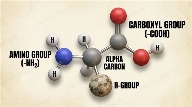
Generalised structure of an amino acid, with the central alpha carbon bonded to an amino group, a carboxyl group, a hydrogen atom and an R-group side chain, all labels pointing to the correct groups
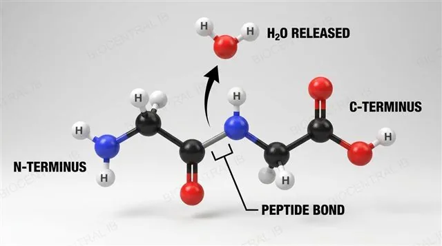
Two amino acids joining by a condensation reaction, releasing one water molecule and forming a peptide bond, with N-terminus, C-terminus and peptide bond labeled
B1.2.2

Peptide bonds: built by condensation

A peptide bond is a covalent C–N bond formed when the carboxyl group of one amino acid reacts with the amino group of another, releasing water.
  • This condensation reaction is the same assembly pattern used across biochemistry; repeating it links many amino acids into a polypeptide.
  • Two amino acids joined this way form a dipeptide; longer chains are polypeptides, a functional polypeptide is a protein.
  • Every polypeptide has direction — a free amino end (N-terminus) and a free carboxyl end (C-terminus).
  • Digestion reverses this by hydrolysis: water is added across each peptide bond to split the chain.
Why it mattersOne reaction — condensation — assembles every protein in every organism, and hydrolysis takes them apart again.
B1.2.3

Some amino acids must come from the diet

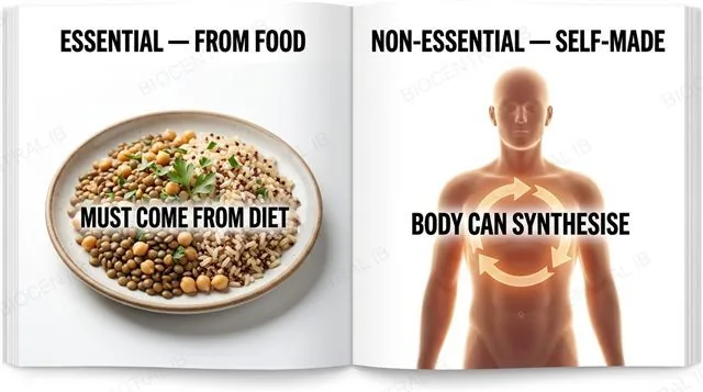
Essential amino acids cannot be synthesised by the human body and must be obtained from food; non-essential amino acids can be made from other amino acids or metabolic intermediates.
  • Roughly half of the 20 standard amino acids are essential for humans; the rest the body can synthesise itself.
  • Vegan diets exclude animal protein, so individual plant foods may be short of one or more essential amino acids.
  • Combining different plant sources — legumes with grains, for example — supplies the full set of essential amino acids across a meal or a day.
Why it matters"Essential" is about dietary need, not importance — every amino acid is used in proteins; the label only tells you whether food must supply it.
Comparison of essential amino acids, which must come from a varied plant-based diet, and non-essential amino acids, which the body can synthesise
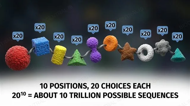
A short peptide chain of amino acid beads, each position marked with a times-20 badge to show that any of the 20 standard amino acids can occupy each position, giving an enormous number of possible sequences
B1.2.4

Twenty amino acids, near-infinite chains

A polypeptide can vary in both length and amino acid order, so the number of possible sequences is enormous.
  • Each position in a chain can be any of the 20 standard amino acids, so a chain of n residues has 20ⁿ possible sequences.
  • A 10-amino-acid chain alone has about 10 trillion possible combinations; real proteins range from tens to thousands of residues.
  • This combinatorial variety lets a small set of amino acids build enzymes, structural fibres, transporters and hormones.
Why it mattersBecause sequence determines folding and folding determines function, the possible range of protein behaviours is effectively unlimited.
B1.2.5

Denaturation: shape lost, sequence intact

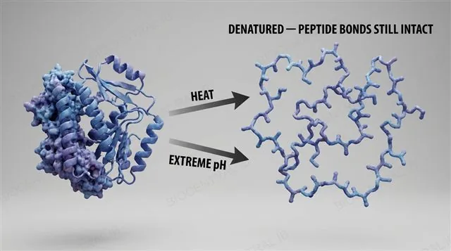
Denaturation is the loss of a protein's three-dimensional shape without breaking the peptide bonds of its primary structure.
  • High temperature disrupts the hydrogen bonds, ionic bonds and hydrophobic interactions that hold the folded shape together.
  • Extreme pH changes the ionisation of acidic and basic R-groups, so their charges change and the same weak interactions break.
  • The unfolded protein can no longer do its job; some proteins refold if conditions return to normal, others do not.
Why it mattersDenaturation is not digestion — hydrolysis breaks peptide bonds, whereas denaturation only unfolds a chain whose sequence is still complete.
A folded functional protein denaturing under heat and a change in pH into an unfolded chain, with peptide bonds remaining intact, as labeled
Quick check

A protein is heated strongly and loses its function, but its amino acid sequence is unchanged. Which bonds were broken, and which remained intact?

Peptide bonds were broken; the weak interactions stayed intact
Hydrogen, ionic and hydrophobic interactions broke; the peptide bonds stayed intact
All bonds were broken, including the peptide bonds
No bonds broke — only the water content changed
Correct answer: the weak interactions broke; the peptide bonds stayed intact. Denaturation disrupts the hydrogen bonds, ionic bonds and hydrophobic interactions that hold the fold, but the covalent peptide bonds of the primary sequence survive.
Part two · HL

Structure and function: from R-group to whole protein

B1.2.6 – B1.2.12
B1.2.6 · HL

R-groups set the chemistry

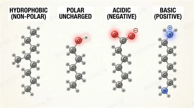
Amino acids are classified by R-group chemistry: hydrophobic (non-polar), hydrophilic polar uncharged, or charged (acidic or basic).
  • Hydrophobic R-groups are non-polar and cluster away from water; polar uncharged R-groups carry –OH or –NH₂ groups that can hydrogen-bond.
  • Charged R-groups are acidic (carboxylate, negative at cell pH) or basic (amino, positive at cell pH).
  • Three special cases: glycine's side chain is a single hydrogen, proline's ring kinks the chain, and two cysteines can form a disulfide bridge.
Why it mattersR-group diversity is the chemical engine of protein form and function — folds, surfaces, active sites and interactions all depend on it.
Classification of amino acid R-groups into non-polar hydrophobic, polar uncharged, and charged acidic or basic, with special cases cysteine, glycine and proline labeled
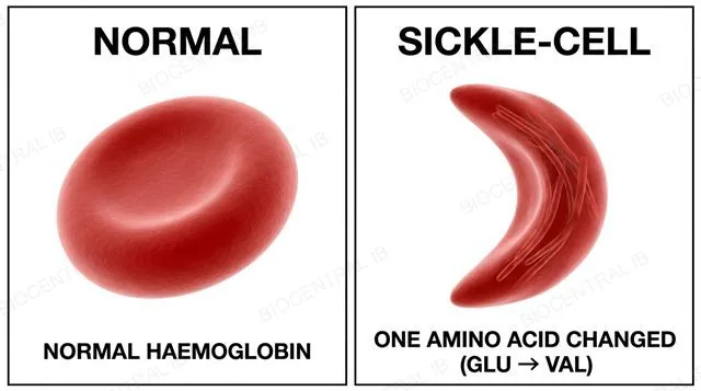
Primary structure as a sequence of amino acid beads determining a folded three-dimensional conformation, with normal and sickled red blood cells showing the effect of one amino acid change from glutamic acid to valine
B1.2.7 · HL

Primary structure sets the fold

Primary structure — the linear amino acid sequence — determines the conformation of the whole protein.
  • The sequence sets which R-groups are available, and therefore where hydrogen bonds, ionic bonds and hydrophobic contacts can form as the chain folds.
  • Sickle-cell disease shows the stakes: one substitution — glutamic acid to valine at position 6 of β-globin — changes a single R-group's chemistry.
  • The altered haemoglobin aggregates into fibres when deoxygenated, distorting red blood cells into sickle shapes and reducing oxygen transport.
Why it mattersOne changed amino acid out of hundreds can reshape and disable a protein — the sequence is information.
B1.2.8 · HL

The α-helix coils the backbone

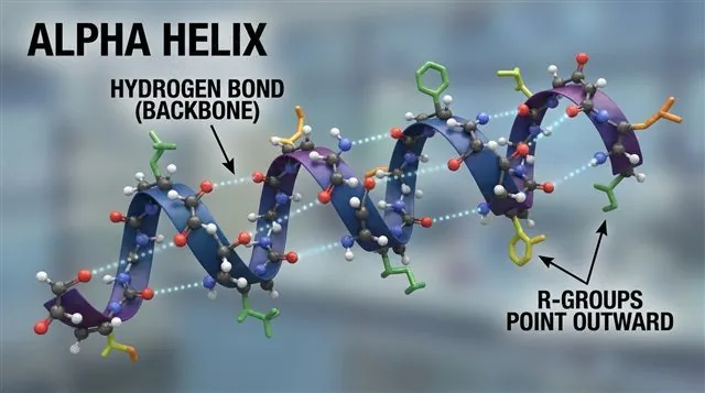
An α-helix is a regular coil of the polypeptide backbone, held together by hydrogen bonds between backbone C=O and N–H groups.
  • Hydrogen bonds link residues about four positions apart along the same chain, twisting it into a right-handed coil that repeats at a constant pitch.
  • R-groups point outward from the helix, away from the hydrogen-bonded backbone, so their interactions belong to tertiary structure, not this one.
Why it mattersA single backbone hydrogen-bonding pattern is enough to build a stable, repeating coil out of almost any sequence.
An alpha helix stabilised by hydrogen bonds between backbone C=O and N-H groups four residues apart, with R-groups pointing outward, labeled
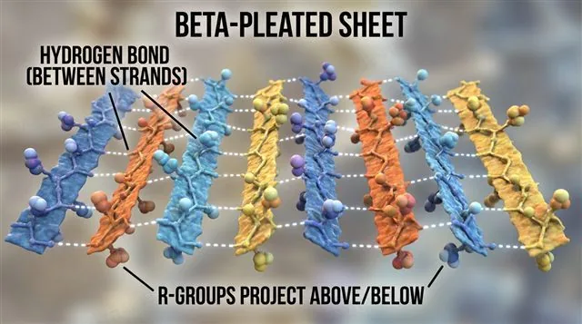
A beta-pleated sheet formed from separate strands lying alongside each other, hydrogen-bonded between strands, labeled
B1.2.8 · HL

The β-pleated sheet bonds between strands

A β-pleated sheet forms when separate sections of the polypeptide chain lie alongside one another and hydrogen-bond between strands rather than within one.
  • The same backbone C=O and N–H hydrogen bonding as the α-helix, but running between adjacent strands instead of along one coiled chain, producing a flatter, pleated geometry.
  • As with the helix, R-groups project above and below the sheet, so their interactions still belong to tertiary structure.
Why it mattersThe same backbone hydrogen bond produces two different geometries — the two recurring structural patterns found in every protein.
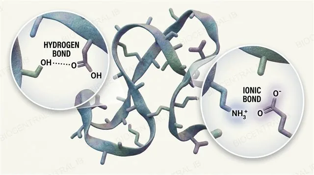
A folded polypeptide with two callouts: a hydrogen bond and an ionic bond, each drawn between the correct R-groups
B1.2.9 · HL

Hydrogen and ionic bonds fold the chain

Tertiary structure is the overall three-dimensional folding of a single polypeptide, and two of its four stabilising interactions are hydrogen bonds and ionic bonds between R-groups.
  • Hydrogen bonds form between polar R-groups wherever a hydrogen-bond donor and acceptor end up close together as the chain folds.
  • Ionic bonds form between oppositely charged R-groups — an acidic side chain's negative charge attracting a basic side chain's positive charge.
Why it mattersBoth interactions are individually weak, but folding brings enough of them together at once to hold a stable shape.
B1.2.9 · HL

Disulfide bridges and hydrophobic interactions lock it in

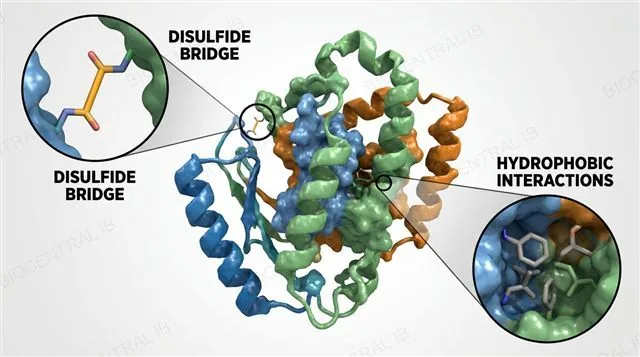
The other two interactions stabilising tertiary structure are disulfide bridges and hydrophobic interactions between R-groups.
  • Disulfide bridges are covalent S–S bonds between two cysteine R-groups — the only covalent bonds, beyond peptide bonds, that hold tertiary structure together, and the strongest of the four.
  • Hydrophobic interactions bury non-polar R-groups in the protein's interior, away from water — not a bond in the same sense, but still a major folding force.
Why it mattersA protein's function depends on this precise 3D shape, and all four interactions together are what keep the chain folded.
A folded polypeptide with two callouts: a disulfide covalent bond between two cysteine R-groups and hydrophobic interactions burying non-polar R-groups in the core
B1.2.10 · HL

Polarity decides what faces water

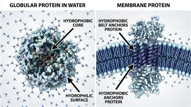
Where each R-group sits in a folded protein is set by its polarity — hydrophobic groups inside, hydrophilic groups outside.
  • In soluble globular proteins, non-polar R-groups cluster in the core while polar and charged R-groups coat the surface.
  • Integral membrane proteins invert the logic: a belt of non-polar R-groups matches the hydrophobic core of the bilayer and anchors the protein.
  • Amino acid composition therefore predicts where a protein lives — cytoplasm, membrane or extracellular matrix.
Why it mattersThe hydrophobic effect — burying non-polar groups — is one of the main thermodynamic drivers of folding itself.
A globular protein with a hydrophobic core and hydrophilic surface, next to a membrane protein whose hydrophobic region anchors it in the phospholipid bilayer
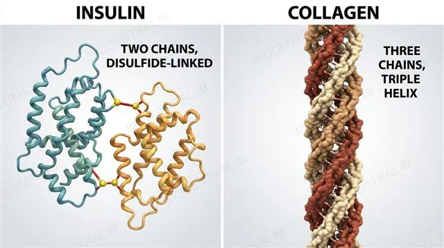
Insulin's two chains joined by disulfide bridges beside collagen's three chains wound into a triple helix, labeled as non-conjugated quaternary proteins
B1.2.11 · HL

Non-conjugated proteins: chains only

Quaternary structure is the association of two or more polypeptide chains into one functional protein; a non-conjugated protein is built from chains alone, with no non-protein group attached.
  • Insulin assembles from two chains joined by disulfide bridges — the same covalent bond type that stabilises tertiary structure, now linking separate chains together instead of one chain to itself.
  • Collagen assembles from three chains wound into a triple helix, giving it the elongated, rope-like structure that underlies its tensile strength.
Why it mattersQuaternary structure can be built entirely from polypeptide chains — no extra chemistry is required to get multiple subunits working as one protein.
B1.2.11 · HL

Haemoglobin: subunits plus a prosthetic group

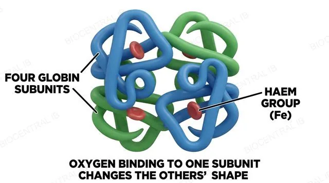
A conjugated protein has a non-protein prosthetic group attached to its polypeptide subunits; haemoglobin is the required example.
  • Haemoglobin assembles four globin subunits, each carrying an iron-containing haem prosthetic group that is what actually binds oxygen.
  • The four subunits cooperate: oxygen binding to one subunit changes the shape of the others, making them bind oxygen more readily in turn — this is what produces the sigmoid oxygen-dissociation curve.
Why it mattersMulti-subunit assembly adds regulation and cooperativity that a single chain cannot achieve on its own.
Haemoglobin's four globin subunits each carrying an iron-containing haem prosthetic group, labeled as a conjugated quaternary protein
B1.2.12 · HL

Globular proteins: compact and dynamic

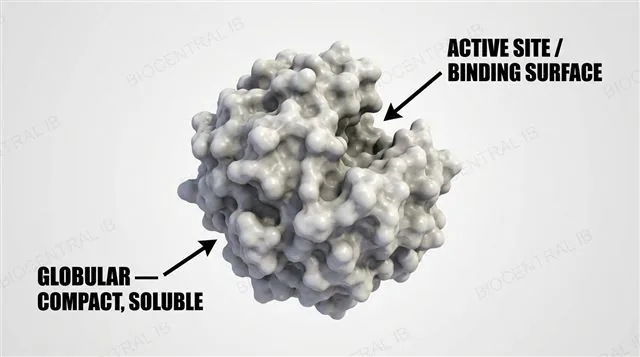
Globular proteins fold into a compact, roughly spherical shape, generally soluble and built for dynamic roles.
  • Enzymes, haemoglobin and insulin are all globular — their folded shape creates active sites, binding pockets and surfaces that other molecules interact with.
  • Being soluble matters functionally: a catalytic, transport or signalling protein has to move through an aqueous environment to reach what it acts on.
Why it mattersA compact fold is what lets a protein present a precise, reusable working surface — the basis of catalysis, transport and signalling alike.
A compact, roughly spherical globular protein such as an enzyme, with a labeled active site or binding surface
An elongated, strand-like fibrous protein such as collagen's triple helix, with its structural role labeled
B1.2.12 · HL

Fibrous proteins: elongated and strong

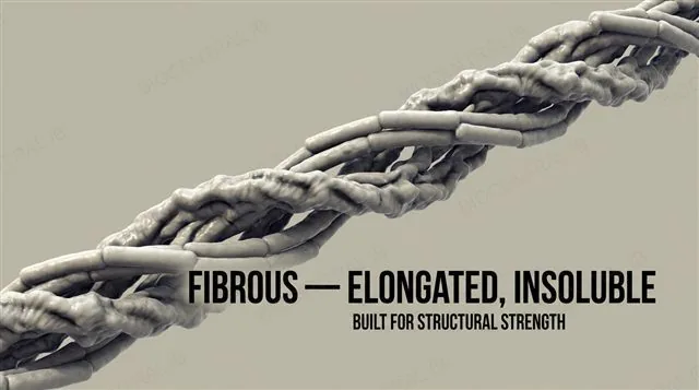
Fibrous proteins are elongated and strand-like, generally insoluble and built for structural roles.
  • Collagen, keratin and silk are all fibrous — their shape trades the compact working surfaces of a globular protein for raw mechanical strength.
  • Collagen's repeating Gly-X-Y sequence and triple helix suit it to load-bearing connective tissue; silk's β-sheets give it exceptional tensile strength for its weight.
Why it mattersProtein shape is not decorative — globular versus fibrous is the direct structural basis of what each protein does.
Quick check · HL

In a water-soluble globular protein, non-polar R-groups are found in the core. What best explains why?

They form peptide bonds with one another
They are attracted to the water outside the protein
Burying them away from water is thermodynamically favourable — the hydrophobic effect
They are pulled into the core by disulfide bridges
Correct answer: the hydrophobic effect. Non-polar R-groups cannot hydrogen-bond with water, so folding buries them in the interior, maximising the entropy of the surrounding water and stabilising the folded protein.

Key vocabulary

Worth being able to define in a single sentence each

Amino acid
Molecule with an alpha carbon bonded to an amino group, a carboxyl group, an H and an R-group.
Peptide bond
Covalent C–N bond between amino acids, formed by condensation.
Polypeptide
A chain of many amino acids linked by peptide bonds.
Essential amino acid
An amino acid the body cannot synthesise; it must come from the diet.
Denaturation
Loss of 3D shape without breaking peptide bonds; caused by heat or pH.
Primary structure HL
The linear amino acid sequence of a polypeptide.
Secondary structure HL
α-helix and β-pleated sheet, held by backbone hydrogen bonds.
Tertiary structure HL
Overall 3D fold of one chain, held by R-group interactions.
Quaternary structure HL
Assembly of two or more polypeptide chains into one protein.
Conjugated protein HL
Protein with a non-protein prosthetic group, e.g. haem in haemoglobin.

Where this shows up again

A1.2 · D1.2
Ribosomes synthesise proteins from amino acids using the genetic code. How does the sequence of bases in DNA (A1.2) ultimately determine the primary structure of a protein?
D1.3 · D3.2
Mutations in DNA (D1.3) can alter a single amino acid in a protein. Using sickle cell disease as an example, explain how a change in primary structure can alter quaternary structure and reduce the protein's ability to perform its function.

B1.2 Proteins — one-page recap

Screenshot this slide to revise from

Amino acids & peptide bonds
  • Backbone: amino + carboxyl + H + R-group on one alpha carbon.
  • Condensation joins amino acids (releases water); hydrolysis breaks them apart.
Diet & sequence variety
  • Essential amino acids must come from food; vegan diets need mixed plant sources.
  • 20 amino acids → 20ⁿ possible sequences; order and length set the protein.
Denaturation (SL)
  • Heat or pH unfolds the protein; weak interactions break.
  • Peptide bonds stay intact — denaturation is not hydrolysis.
Primary & secondary HL
  • Primary sequence determines the fold (sickle cell: Glu→Val).
  • Secondary: α-helix and β-sheet held by backbone C=O···N–H hydrogen bonds.
Tertiary structure HL
  • R-group interactions: hydrogen, ionic, disulfide bridges, hydrophobic core.
  • Polarity places hydrophobic groups inside, hydrophilic groups outside.
Quaternary & form HL
  • Subunits: insulin (non-conjugated), haemoglobin (conjugated, haem).
  • Globular (soluble, dynamic) vs fibrous (insoluble, structural).

Sequence becomes shape becomes function

Twenty amino acids, arranged and folded, build every enzyme, fibre and signal in the living world.
B1.2 Proteins · BioCentral IB
Use ↓ ↑ or click to navigate
01 / 22