BioCentral IBBioCentral IB
IB Biology · Theme B · B2.1

Membranes and membrane transport

How a two-molecule-thick sheet of lipids becomes a selective gatekeeper — letting oxygen slip through, holding ions back, and powering the pumps that make nerve impulses possible.
Guiding questions

How do molecules of lipid and protein assemble into biological membranes?

What determines whether a substance can pass through a biological membrane?

Part one

The membrane: structure and selective permeability

B2.1.1 – B2.1.10
B2.1.1

Lipid bilayers build themselves

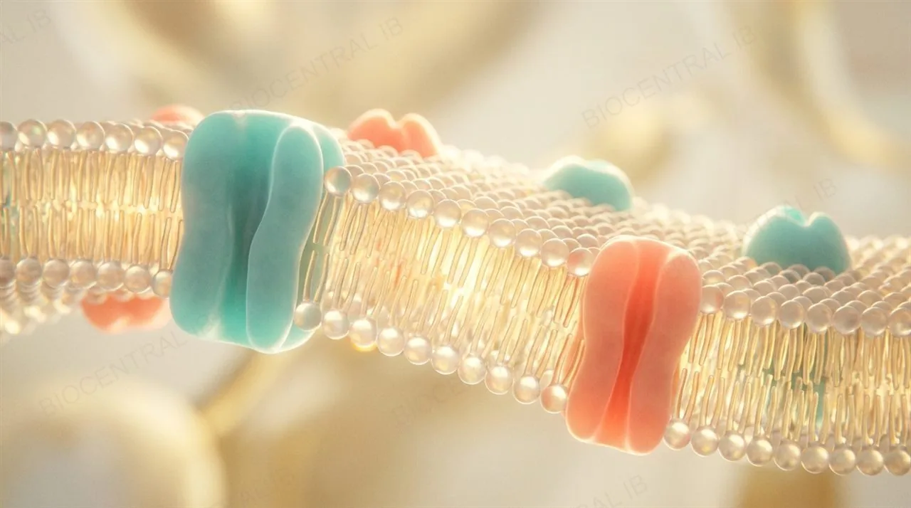
A phospholipid bilayer is two layers of phospholipids arranged tail-to-tail — the structural basis of every cell membrane.
  • Each phospholipid is amphipathic: a hydrophilic phosphate head faces the water on either side, while the two hydrophobic fatty acid tails face each other in the middle.
  • In water, phospholipids spontaneously assemble tails-inward because this minimises contact between the hydrophobic tails and water.
  • The arrangement is self-sealing — a small tear reseals itself for the same reason, which is why a membrane can fuse, bend and pinch off without leaking.
Why it mattersNo enzyme builds the bilayer — the hydrophobic effect alone assembles it, which is why membranes form so readily in cells.
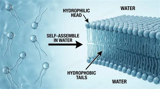
Phospholipids scattered in water self-assembling into a continuous bilayer with hydrophilic heads facing the water and hydrophobic tails meeting tail-to-tail, labeled
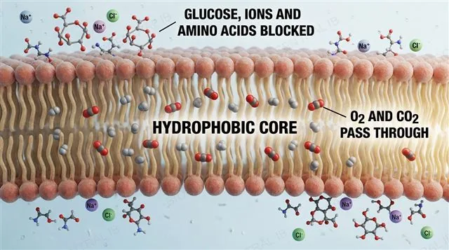
A phospholipid bilayer acting as a barrier: small non-polar oxygen and carbon dioxide pass through the hydrophobic core while glucose, amino acids and ions are blocked, labeled
B2.1.2

The hydrophobic core is a barrier

The hydrophobic hydrocarbon core of the bilayer has very low permeability to large molecules and hydrophilic particles, including ions and polar molecules.
  • Moving a charged or polar particle through a non-polar environment is energetically very unfavourable, so ions such as Na⁺, K⁺ and Cl⁻ cannot cross directly.
  • Larger polar molecules such as glucose and amino acids are also excluded for the same reason.
  • This is what makes the membrane useful: an impermeable barrier to most solutes means the cell can control entry and exit by choosing which transport proteins to place in the membrane.
Why it mattersIf every solute crossed freely, cells could never build an internal environment different from their surroundings.
B2.1.3

Simple diffusion: the free route

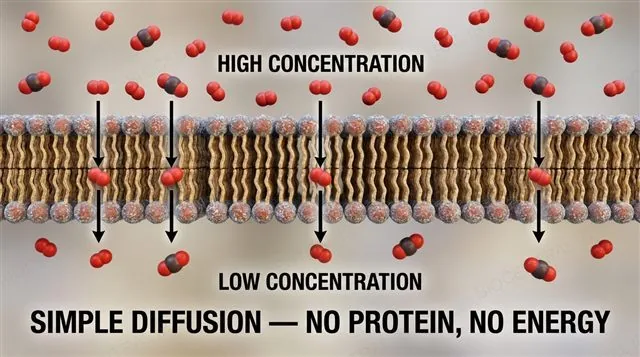
Simple diffusion is the net movement of small non-polar particles down their concentration gradient, directly through the bilayer — no protein, no energy.
  • Small non-polar molecules such as oxygen (O₂), carbon dioxide (CO₂) and nitrogen (N₂) dissolve readily in the hydrophobic core and cross freely.
  • Very small polar molecules such as water cross slowly by this route too, though most water movement uses aquaporins instead.
  • The rate of simple diffusion is proportional to the concentration gradient and never saturates — there are no limited binding sites to fill, unlike protein-mediated transport.
Why it mattersRespiration and photosynthesis depend on O₂ and CO₂ crossing membranes freely down their gradients.
Oxygen and carbon dioxide diffusing down their concentration gradient straight through a phospholipid bilayer, with no protein and no energy, labeled
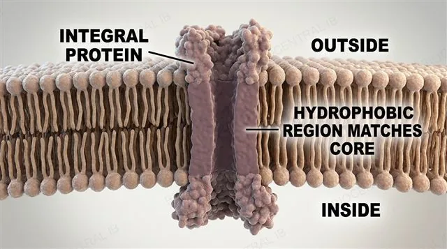
An integral protein spanning completely through the phospholipid bilayer, with hydrophobic amino acids matching the hydrophobic core, labeled
B2.1.4

Integral proteins span the bilayer

Integral proteins are embedded in one or both lipid layers of the membrane.
  • Many integral proteins are transmembrane — they span the bilayer completely, with a band of hydrophobic amino acids matching the hydrophobic core so the protein sits stably in the membrane rather than being expelled from it.
  • Because they cross the whole membrane, transmembrane proteins are what make transport possible — a channel or pump has to reach both sides at once to move anything across.
Why it mattersA protein's amino acid sequence, not just its shape, decides whether it can sit inside a hydrophobic membrane core at all.
B2.1.4

Peripheral proteins stay on the surface

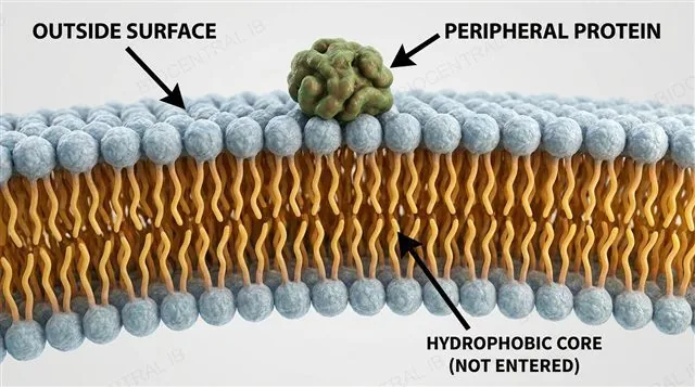
Peripheral proteins are attached to one or other surface of the bilayer, without entering the hydrophobic core.
  • They sit on the surface, often bound to an integral protein or to the phospholipid heads themselves, held on by weaker interactions than the ones anchoring an integral protein in place.
  • Between them, integral and peripheral proteins carry out most of a membrane's jobs: transport, enzymatic catalysis, receptor signalling and cell adhesion — their structures are as diverse as those jobs.
Why it mattersDifferent membranes hold different proteins, which is precisely what makes each membrane selective for its own cell's needs.
A peripheral protein attached to the outer surface of the phospholipid bilayer, not entering the hydrophobic core, labeled
B2.1.5

Osmosis: water follows solute

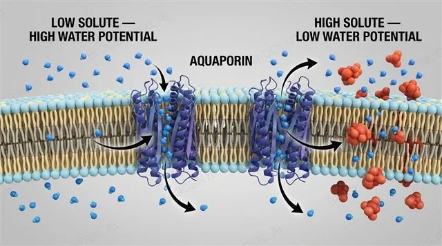
Osmosis is the net movement of water across a partially permeable membrane from lower solute concentration (higher water potential) to higher solute concentration (lower water potential).
  • Water moves by random motion, but because the membrane is impermeable to the solute, the net flow is toward the side with more solute — toward lower water potential.
  • Aquaporins are channel proteins specific to water; they greatly increase a membrane's water permeability wherever fast water movement is needed, such as kidney tubules and plant roots.
  • Osmosis is passive — no energy is used; only the water-potential gradient drives it.
Why it mattersOsmosis sets cell volume: animal cells burst or shrivel when water moves, while plant cells rely on their cell wall to become turgid instead.
Water molecules passing through aquaporin channels from a low-solute to a high-solute side, while solute particles are too large to pass, labeled
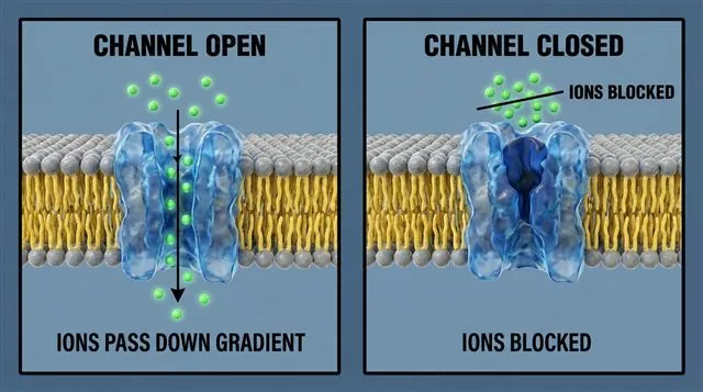
A channel protein open and letting ions pass down their gradient, compared with the same channel closed and blocking the ions, labeled
B2.1.6

Channels: selective doors for facilitated diffusion

Facilitated diffusion moves charged or larger particles down their concentration gradient through a transport protein — still passive, still no energy.
  • A channel protein forms a water-filled pore through the membrane, often gated, allowing rapid passive flow of a specific ion or small molecule.
  • The channel's structure makes the membrane selectively permeable: the right ion passes when the channel is open, and nothing passes when it is closed.
  • Facilitated diffusion shows saturation — once every transport protein is occupied, the rate plateaus, unlike simple diffusion.
Why it mattersGated channels let a cell switch specific ion flows on and off — the foundation of nerve signalling.
B2.1.7

Pumps: uphill, using ATP

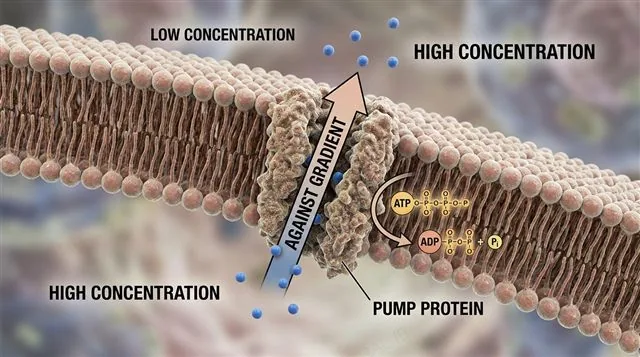
Active transport moves particles against their concentration gradient using energy, typically from ATP hydrolysis, and always needs a carrier-type pump protein.
  • A pump uses energy from ATP to transfer specific particles across a membrane, so it can move them from low to high concentration.
  • Primary active transport couples ATP hydrolysis directly to the uphill movement — the sodium–potassium pump is the classic example.
  • Secondary active transport instead uses an ion gradient, itself built by primary active transport, to drive a second solute uphill.
Why it mattersPumps build the gradients that make everything else — nerve impulses, nutrient uptake, water balance — possible.
A pump protein using ATP to move a particle from low to high concentration, against the concentration gradient, labeled
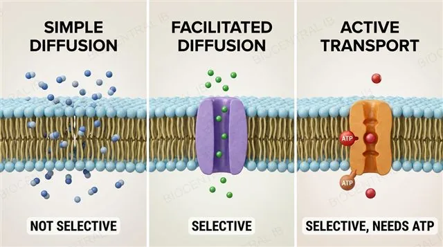
Three transport routes compared: simple diffusion through the bilayer, facilitated diffusion through a channel, and active transport by a pump using ATP, labeled
B2.1.8

Permeability is selective, not uniform

Facilitated diffusion and active transport make membranes selectively permeable; simple diffusion is not selective — it depends only on size and polarity.
  • Which route a substance takes — free diffusion, a channel, a carrier or a pump — depends on its size, polarity and charge, and on which transport proteins that membrane contains.
  • Simple diffusion lets any sufficiently small non-polar particle through; it cannot discriminate between one such molecule and another.
  • Because different cell types express different combinations of channels and pumps, a neuron, a kidney cell and a muscle cell build very different internal environments from the same extracellular fluid.
Why it mattersSelectivity is what turns a simple lipid sheet into a precisely controlled cellular boundary.
B2.1.9

Sugar tags on the outside

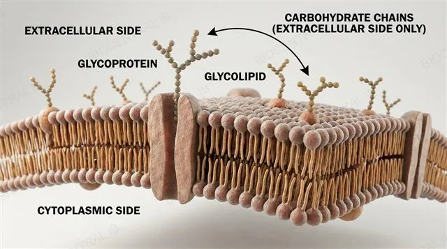
Glycoproteins and glycolipids are membrane proteins or lipids with carbohydrate chains attached, found on the extracellular side of the membrane.
  • The carbohydrate chains always project into the extracellular space, never into the cytoplasm.
  • These sugar chains act as identification tags for cell–cell recognition — the immune system uses them to distinguish self from non-self.
  • They form the basis of the ABO blood-group antigens and help cells of the same tissue recognise and adhere to one another.
Why it mattersThe molecular "name tags" on a cell's surface decide whether immune cells and neighbouring cells treat it as friend or foe.
A glycoprotein and a glycolipid in a membrane, with carbohydrate chains projecting into the extracellular side, labeled with their recognition and adhesion roles
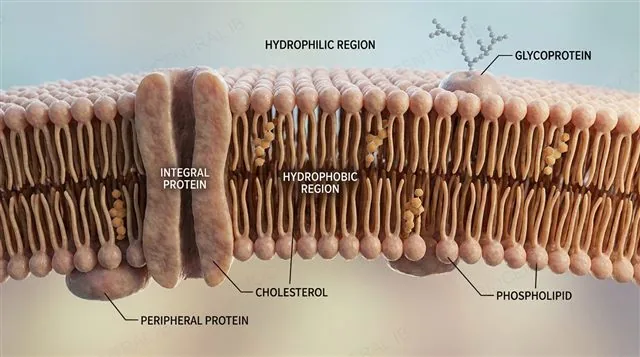
The fluid mosaic model: a phospholipid bilayer with integral, peripheral and glycoproteins plus cholesterol, with hydrophilic and hydrophobic regions labeled
B2.1.10

Fluid mosaic: a moving, mixed mosaic

The fluid mosaic model describes the membrane as a dynamic structure: phospholipids and proteins drift laterally within their layer, like tiles in a mosaic.
  • A 2D drawing of the model must show phospholipids, integral and peripheral proteins, glycoproteins and cholesterol, with hydrophilic and hydrophobic regions indicated.
  • "Fluid" refers to lateral movement: phospholipids and many proteins diffuse sideways within their leaflet rather than staying fixed.
  • Frye and Edidin's cell-fusion experiment showed this directly — fluorescent mouse and human membrane proteins mixed completely across a fused cell within about 40 minutes.
Why it mattersMembrane fluidity is what lets membranes bend, fuse, and pinch off — and it is exactly what the HL section turns to next.
Quick check

A membrane separates two solutions and contains only phospholipids — no proteins. Which substance crosses it most easily by simple diffusion?

Na⁺ ions
Oxygen (O₂)
Glucose
An amino acid
Correct answer: oxygen. O₂ is small and non-polar, so it dissolves in the hydrophobic core and crosses freely. Ions are charged, and glucose and amino acids are large and polar — all are blocked by the core and need transport proteins instead.
Part two · HL

Membrane fluidity and specialised transport

B2.1.11 – B2.1.17
B2.1.11 · HL

Kinks keep membranes fluid

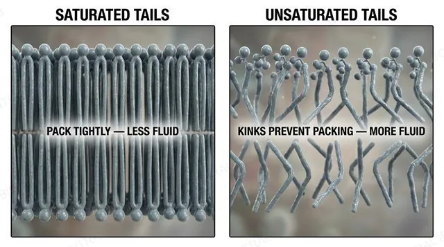
Unsaturated fatty acid tails have kinks at their C=C double bonds, so they pack less tightly and make membranes more fluid at a given temperature.
  • Saturated tails are straight and pack closely, which raises the membrane's melting point and makes it stronger at higher temperatures.
  • Unsaturated tails cannot pack as tightly, so membranes rich in them stay fluid and flexible at lower temperatures.
  • Cold-water fish and cold-tolerant plants increase the proportion of unsaturated fatty acids in their membranes — a compensation called homeoviscous adaptation.
Why it mattersAn organism can tune its membranes to its habitat simply by changing the mix of saturated and unsaturated tails.
Saturated straight phospholipid tails packing tightly versus unsaturated kinked tails packing loosely, with fluidity labels
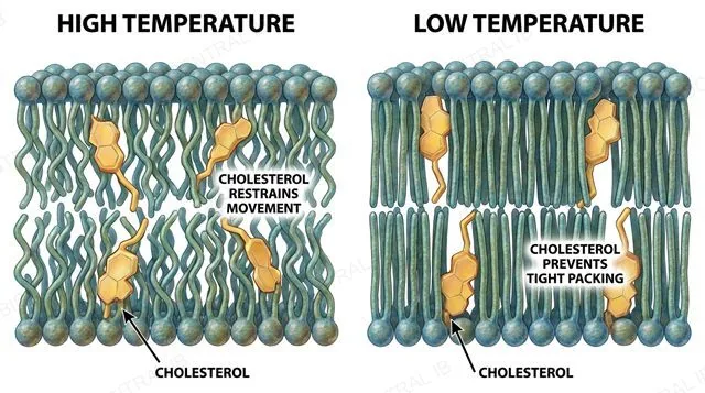
Cholesterol wedged between phospholipid tails, restraining movement at high temperature and preventing stiffening at low temperature, labeled
B2.1.12 · HL

Cholesterol: the fluidity buffer

In animal cells, cholesterol sits between phospholipid tails and modulates membrane fluidity — stabilising at high temperatures, preventing stiffening at low ones.
  • At high temperature, cholesterol restrains phospholipid movement, so the membrane does not become excessively fluid.
  • At low temperature, it stops the tails from packing too closely, so the membrane does not solidify.
  • Plant cells lack cholesterol and use other sterols for the same buffering role.
Why it mattersCholesterol widens the temperature range over which an animal membrane keeps the right consistency.
B2.1.13 · HL

Endocytosis brings material in

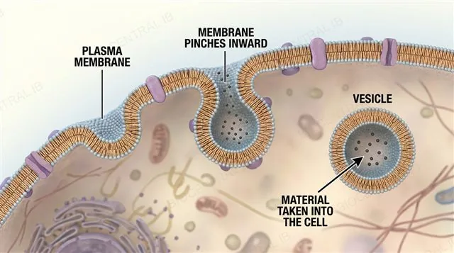
Endocytosis brings material into the cell as the membrane pinches inward to form a vesicle — a process only a fluid membrane can perform.
  • The bilayer bends, pinches off and reseals around the internalised material without ever breaking apart — fluidity is what lets it deform this much and still stay intact.
  • Endocytosis covers three related routes: phagocytosis (engulfing large particles), pinocytosis (taking in extracellular fluid) and receptor-mediated endocytosis (concentrating one target molecule before internalising it).
Why it mattersA rigid membrane could never eat or drink — every route in requires the bilayer to physically fold in on itself.
A cell membrane pinching inward around extracellular material to form an internalised vesicle, labeled
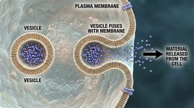
A vesicle fusing with the plasma membrane from inside the cell and releasing its contents outside, labeled
B2.1.13 · HL

Exocytosis sends material out

Exocytosis releases material from the cell as an internal vesicle travels to the plasma membrane and fuses with it.
  • Once the vesicle membrane merges with the plasma membrane, its contents are released directly into the extracellular space — the reverse geometry of endocytosis, but the same fluidity requirement.
  • This is how cells secrete products such as digestive enzymes or hormones; both endocytosis and exocytosis require energy to drive the membrane rearrangement.
Why it mattersSecretion has no other route out — anything a cell exports in bulk leaves through exocytosis.
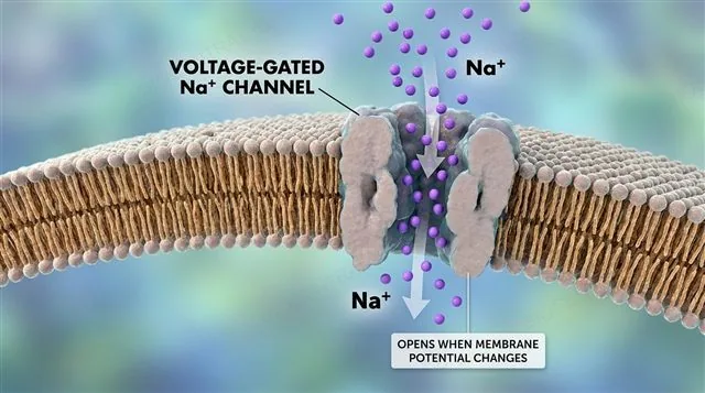
A voltage-gated sodium channel in a neuron's axon opening in response to a change in membrane potential, with Na+ flowing in, labeled
B2.1.14 · HL

Voltage-gated channels sense membrane potential

A voltage-gated ion channel opens in response to a change in membrane potential, not to any chemical binding.
  • Sodium and potassium channels in a neuron's axon are voltage-gated — each one opens once the membrane depolarises past its own threshold, then closes again.
  • Their opening and closing in strict sequence along the axon is what underlies the action potential, propagating the electrical signal down the neuron.
Why it mattersA voltage-gated channel needs no messenger molecule at all — the electrical signal itself is what opens the gate.
B2.1.14 · HL

Ligand-gated channels sense a chemical signal

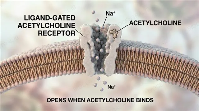
A ligand-gated ion channel opens only when a specific chemical signal binds to it.
  • The nicotinic acetylcholine receptor is the classic example: when acetylcholine binds at a synapse, the channel opens and Na⁺ flows in, depolarising the postsynaptic membrane.
  • Unlike a voltage-gated channel, this one responds to a messenger molecule released by another cell, not to the membrane's own electrical state.
Why it mattersBetween the two gating mechanisms, a neuron can convert both electrical and chemical signals into the same currency: an ion flow.
A ligand-gated acetylcholine receptor channel at a synapse opening when acetylcholine binds, with Na+ flowing in, labeled
B2.1.15 · HL

The sodium–potassium pump: an exchange transporter

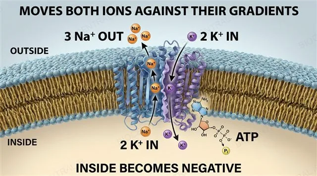
The Na⁺/K⁺-ATPase uses one ATP per cycle to pump 3 Na⁺ out and 2 K⁺ in, both against their concentration gradients.
  • Because it exchanges one kind of particle for another while moving both uphill, it is an exchange transporter and the clearest example of primary active transport.
  • The pump is electrogenic: each cycle exports one more positive charge than it imports, directly contributing to the inside-negative membrane potential.
  • The Na⁺ gradient it builds is also the energy source for secondary active transport, such as the glucose symporter.
Why it mattersThis single pump underlies resting membrane potential in neurons and powers nutrient uptake across the body.
The sodium-potassium pump exchanging 3 Na+ out for 2 K+ in per ATP, against both gradients, with the electrogenic effect labeled
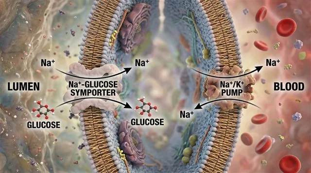
A sodium-dependent glucose symporter using the Na+ gradient to pull glucose into an intestinal epithelial cell, with the Na+/K+ pump maintaining the gradient, labeled
B2.1.16 · HL

Cotransport: glucose rides the Na⁺ gradient

Sodium-dependent glucose cotransport is indirect active transport: glucose is pulled against its gradient by coupling its uptake to Na⁺ flowing down its gradient.
  • The symporter uses the Na⁺ gradient already established by the Na⁺/K⁺ pump — it does not use ATP directly.
  • In the small intestine and the kidney's proximal tubule, this lets cells absorb glucose even when its own gradient would otherwise stop net uptake.
  • The Na⁺/K⁺ pump keeps the Na⁺ gradient steep by continually exporting Na⁺, so the cotransporter keeps working.
Why it mattersIt explains how the gut and kidney scavenge every last glucose molecule from the fluid passing through them.
B2.1.17 · HL

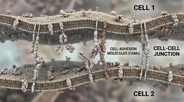
Cell-adhesion molecules build tissues

Cell-adhesion molecules (CAMs) are membrane proteins that bind cells together; different forms of CAM are used for different types of cell–cell junction.
  • CAMs on one cell bind matching CAMs or other molecules on a neighbouring cell, holding the two membranes together.
  • Different tissues express different CAMs, which is how cells of the same tissue recognise and stick to one another rather than to unrelated cells.
  • This selective adhesion is what assembles individual cells into structured tissues and organs.
Why it mattersWithout CAMs, multicellular life would be a loose pile of cells, not an organised body.
Two cells joined by matching cell-adhesion molecules forming a cell-cell junction, labeled
Quick check · HL

The sodium–potassium pump moves 3 Na⁺ out and 2 K⁺ in per ATP. Why does this make the inside of the cell negative?

It moves equal numbers of charges, so the potential does not change
K⁺ carries a negative charge
Each cycle exports one more positive charge than it imports, so the inside loses net positive charge
ATP hydrolysis releases electrons into the cytoplasm
Correct answer: each cycle exports one more positive charge than it imports. Three Na⁺ leave for every two K⁺ entering, so the cytoplasm loses one net positive charge per cycle — the pump is electrogenic and contributes directly to the inside-negative resting potential.

Key vocabulary

Worth being able to define in a single sentence each

Phospholipid bilayer
Two layers of phospholipids, tails inward — the basis of cell membranes.
Amphipathic
Having both a hydrophilic and a hydrophobic region, like a phospholipid.
Simple diffusion
Net movement of small non-polar particles down their gradient through the bilayer.
Osmosis
Net movement of water across a partially permeable membrane toward higher solute concentration.
Facilitated diffusion
Passive movement down a gradient through a channel or carrier protein.
Active transport
Movement against a gradient by a pump, using energy such as ATP.
Glycoprotein / glycolipid
Membrane protein or lipid with a carbohydrate chain, used in recognition.
Fluid mosaic model
Membrane as a fluid bilayer with a mosaic of proteins drifting laterally.
Cholesterol HL
Steroid between phospholipid tails that buffers membrane fluidity in animal cells.
Endo-/exocytosis HL
Vesicle formation and fusion that move materials into and out of cells.
Gated ion channel HL
Channel opened by a voltage change or by a chemical signal binding.
Cell-adhesion molecule HL
Membrane protein that binds cells together at cell–cell junctions.

Where this shows up again

B1.1
Phospholipids are the structural foundation of cell membranes. How do the properties of phospholipids, introduced in B1.1, enable the spontaneous formation of bilayers in aqueous environments?
B2.2
Membrane proteins are synthesised by ribosomes on the rough endoplasmic reticulum. How does the endomembrane system (B2.2) deliver these proteins to their correct membrane destinations?
C2.2
The sodium-potassium pump maintains resting membrane potential, which is essential for nerve impulse transmission (C2.2). Explain how the pump's unequal transport ratio (3 Na⁺ out : 2 K⁺ in) contributes to the inside-negative membrane potential.
D2.3
Water potential in plant tissues (D2.3) depends on both solute concentration and pressure. How does osmosis, driven by differences in water potential, explain the movement of water from soil into root hair cells and then through the root to the xylem?

B2.1 Membranes and membrane transport — one-page recap

Screenshot this slide to revise from

Bilayer & barrier
  • Amphipathic phospholipids self-assemble tails-inward in water.
  • Hydrophobic core blocks ions, polar and large molecules.
Passive movement
  • Simple diffusion: O₂/CO₂ through the bilayer, down gradient.
  • Facilitated diffusion: channels/carriers, down gradient, saturates.
Osmosis & active transport
  • Osmosis: water toward higher solute via aquaporins.
  • Pumps: ATP moves particles against their gradient.
Membrane parts
  • Integral (embedded) vs peripheral (surface) proteins.
  • Glycoproteins/glycolipids tag the extracellular surface.
Fluidity HL
  • Unsaturated tails (kinks) increase fluidity; saturated pack tight.
  • Cholesterol buffers fluidity high and low temperatures.
Specialised transport HL
  • Na⁺/K⁺ pump (3 out : 2 in) is electrogenic; powers cotransport.
  • Gated channels fire neurons; CAMs build tissues.

Two layers of lipids, a world of decisions

Every substance a cell takes in or sends out crosses the same fluid, selective boundary.
B2.1 Membranes and membrane transport · BioCentral IB
Use ↓ ↑ or click to navigate
01 / 22