BioCentral IBBioCentral IB
IB BIOLOGY · THEME B · B2.2

Organelles and compartmentalisation

Why walling a cell into separate rooms lets incompatible chemistry run at once — and how a nucleus, a mitochondrion and a lysosome each get to keep their own rules.
Guiding questions

How are organelles in cells adapted to their functions?

What are the advantages of compartmentalization in cells?

Part one

Cells built from separate working parts

B2.2.1 – B2.2.3
B2.2.1

A cell is built from discrete parts

An organelle is a discrete, membrane-bound subunit within a cell, adapted to perform one specific function.
  • Not every structure counts as an organelle: the cell wall, cytoskeleton and cytoplasm are excluded, since they aren't membrane-bound discrete subunits. The nucleus, vesicles, ribosomes and the plasma membrane are all organelles.
  • Each major organelle has its own job — nucleus (genetic material, control), mitochondrion (aerobic respiration), ribosome (protein synthesis), ER (synthesis/transport), Golgi apparatus (modifies, sorts, packages), lysosome (digestion), vacuole (storage, turgor), chloroplast (photosynthesis, plant cells).
Why it mattersStudying what a single organelle actually does only became possible once ultracentrifuges and cell-fractionation methods existed — progress here followed the tools, not the other way round.
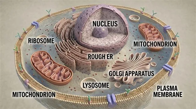
A generalized animal cell with major organelles labeled — nucleus, mitochondrion, ribosome, rough ER, Golgi apparatus, lysosome, plasma membrane
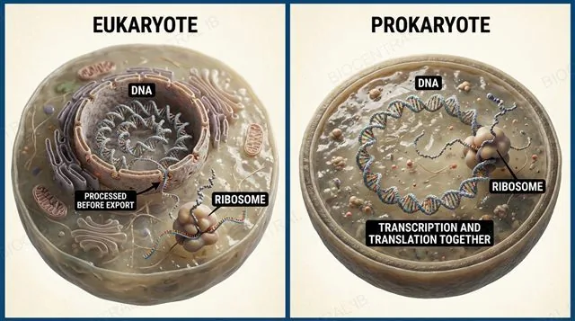
The nuclear envelope separating transcription in the nucleus from translation in the cytoplasm, with mRNA processing shown before export, contrasted with a prokaryote where both happen together
B2.2.2

Separating the nucleus buys editing time

The nuclear envelope separates transcription, in the nucleus, from translation, in the cytoplasm.
  • That separation gives mRNA time to be processed before ribosomes ever see it: introns are spliced out, and a 5′ cap and 3′ poly-A tail are added to protect it and help it exit the nucleus.
  • Prokaryotes have no nuclear envelope, so transcription and translation happen in the same compartment — a ribosome can start translating an mRNA molecule while it is still being transcribed.
Why it mattersWithout this separation, ribosomes could translate unprocessed mRNA — introns and all — before it was ever edited.
B2.2.3

Lysosomes keep dangerous enzymes contained

Compartmentalisation concentrates specific enzymes and substrates together and separates incompatible biochemical processes, each kept at its own optimal conditions.
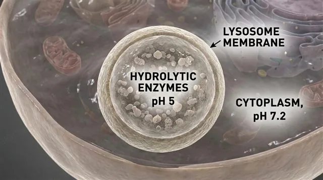
  • A lysosome holds hydrolytic enzymes that work best at an acidic pH of around 5 — sealed away from the near-neutral cytoplasm, at pH ~7.2, where those same enzymes would be far less active.
  • Keeping the two environments separate is what prevents the cell from digesting its own cytoplasmic proteins with its own waste-disposal enzymes.
Why it mattersConcentrating an enzyme with its substrate — and away from everything it shouldn't touch — is what makes a reaction fast and safe at the same time.
A lysosome containing hydrolytic enzymes at pH 5, sealed off from the near-neutral cytoplasm at pH 7.2, labeled
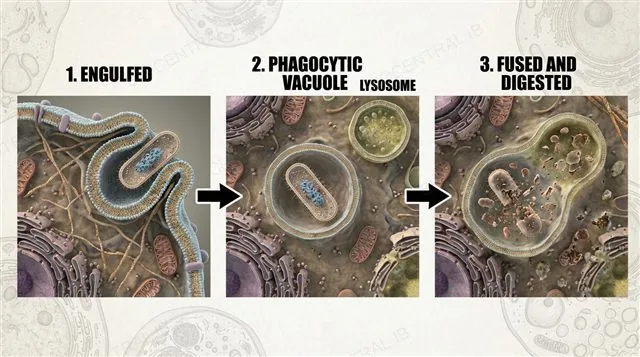
A phagocytic vacuole engulfing a bacterium then fusing with a lysosome to digest it, labeled
B2.2.3

Phagocytic vacuoles isolate what they engulf

A phagocytic vacuole engulfs a pathogen or debris, isolating it from the rest of the cell the instant it's taken in.
  • The vacuole then fuses with a lysosome, delivering the target directly into the acidic, enzyme-rich environment that digests it.
  • Because the fusion happens membrane-to-membrane, the lysosome's hydrolytic enzymes never spill into the general cytoplasm during the process.
Why it mattersDigestion happens exactly where it's needed and nowhere else — the same compartmentalisation principle as B2.2.3's lysosome, applied to a second, real example.
Quick check

A lysosome's enzymes work best at pH 5. Why doesn't the lysosome digest the rest of the cell?

The enzymes are inactive until they leave the lysosome
The cytoplasm is also kept at pH 5
The lysosome's membrane keeps its acidic interior separate from the near-neutral cytoplasm
Lysosomal enzymes cannot digest proteins
Correct answer: the lysosome's membrane keeps its acidic interior separate. The enzymes are fully active hydrolytic enzymes — what protects the rest of the cell is compartmentalisation, not a difference in the enzymes themselves. The cytoplasm sits at a near-neutral pH of ~7.2, where those same enzymes barely function.
Part two · HL

Structure built for a single job

B2.2.4 – B2.2.9
B2.2.4 · HL

The mitochondrion is built for ATP

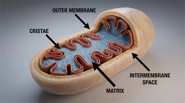
Every structural feature of a mitochondrion increases its capacity to produce ATP by aerobic respiration.
  • A double membrane keeps the intermembrane space small, concentrating the H⁺ gradient built during oxidative phosphorylation; the inner membrane folds into cristae, massively increasing the surface area available for the electron transport chain and ATP synthase.
  • The matrix holds the Krebs cycle's enzymes, kept apart from the cytoplasm where glycolysis runs — each pathway gets its own conditions. Mitochondria carry their own DNA and 70S ribosomes, distinct from the cell's nuclear DNA and 80S cytoplasmic ribosomes.
Why it mattersMitochondria are not part of the endomembrane system — no vesicle traffic connects them to the ER or Golgi. They're semi-autonomous, with their own genome.
A mitochondrion cutaway showing the outer membrane, inner membrane folded into cristae, narrow intermembrane space, and the matrix, labeled
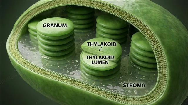
A chloroplast cutaway showing thylakoids stacked into grana with a small internal lumen, and the surrounding stroma, labeled
B2.2.5 · HL

The chloroplast mirrors the same logic

A chloroplast's internal structure separates the light-dependent reactions from the Calvin cycle, just as the mitochondrion separates the electron transport chain from the Krebs cycle.
  • Thylakoid membranes stack into grana, giving a large surface area for the photosystems of the light-dependent reactions; the thylakoid lumen is kept small in volume, concentrating protons for chemiosmosis.
  • The stroma surrounds the thylakoids and holds the Calvin cycle's enzymes, kept separate from the thylakoid space so each set of reactions gets its own pH and enzyme conditions. Chloroplasts also carry their own DNA and 70S ribosomes.
Why it mattersSame principle, two organelles: a small internal volume concentrates a proton gradient, and a large membrane surface area maximises the reactions that build it.
B2.2.6 · HL

Nuclear pores let the nucleus stay in touch

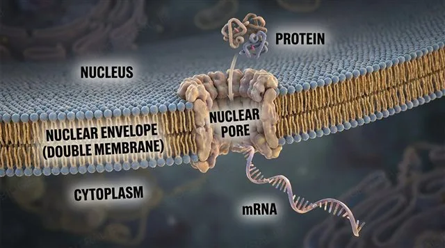
The nuclear envelope is a double membrane punctuated by nuclear pores that regulate exactly what crosses it.
  • Pores let mRNA exit and proteins such as transcription factors enter, rather than allowing free diffusion — the nucleus stays chemically separate from the cytoplasm while still communicating with it.
  • During mitosis and meiosis, the entire nuclear envelope breaks down into vesicles, then reforms around each daughter nucleus once division is complete.
Why it mattersA double membrane with selective pores gives the best of both worlds: real separation, without cutting the nucleus off from the rest of the cell.
The nuclear double membrane with nuclear pores regulating mRNA exiting and proteins entering, labeled
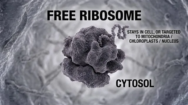
A free ribosome in the cytosol making a protein that stays in the cell or is targeted to mitochondria, chloroplasts or the nucleus, labeled
B2.2.7 · HL

Free ribosomes serve the cytosol

Free ribosomes float unattached in the cytosol, making proteins that mostly stay inside the cell.
  • Proteins made on free ribosomes either remain in the cytosol to do their job there, or are targeted after synthesis to mitochondria, chloroplasts or the nucleus.
  • A signal sequence on the growing protein — not the ribosome itself — is what decides its eventual destination; the ribosome is otherwise identical to a membrane-bound one.
Why it mattersNot every ribosome sits on the ER — free ribosomes are just as real, and just as busy, making a different set of proteins entirely.
B2.2.7 · HL

Membrane-bound ribosomes serve export

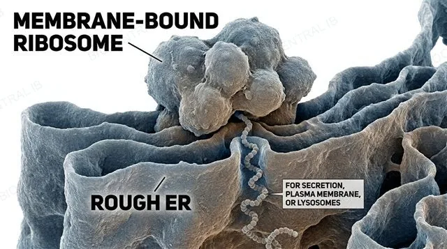
Membrane-bound ribosomes sit on the rough ER, feeding their protein directly into the endomembrane system as it's made.
  • These ribosomes make proteins destined for secretion out of the cell, insertion into the plasma membrane, or delivery to a lysosome — all three routes start the same way, threading the growing chain into the ER lumen.
  • The same signal sequence that keeps a protein on a free ribosome sends this one to the ER instead — one shared mechanism deciding two very different fates.
Why it mattersAnything the cell secretes or displays on its surface has to pass through this route first — there's no other way out.
A membrane-bound ribosome on the rough ER feeding a growing protein into the ER lumen, destined for secretion, labeled
B2.2.8 · HL

The Golgi finishes what ribosomes started

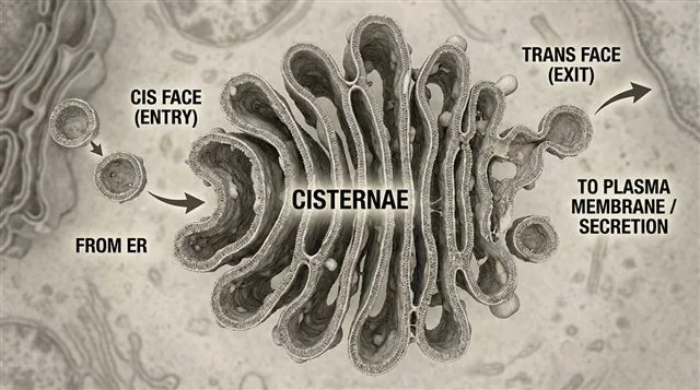
The Golgi apparatus is a stack of flattened membrane sacs, cisternae, with a distinct entry face and exit face, that processes and secretes proteins.
  • Vesicles from the ER arrive at the cis (entry) face; as proteins and lipids pass through the stack they're modified — glycosylation, the addition of sugar chains, is one example.
  • Finished molecules are sorted and packaged into new vesicles that bud off from the trans (exit) face, headed for secretion, the plasma membrane, or lysosomes.
Why it mattersThe Golgi doesn't synthesise proteins — ribosomes already did that. Its job is modifying, sorting and packaging what arrives.
The Golgi apparatus as stacked cisternae receiving vesicles at the cis face and budding new vesicles from the trans face, labeled
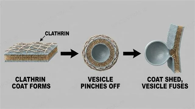
A clathrin lattice coating a membrane and pinching off a vesicle, then the coat detaching before the vesicle fuses with its target membrane, labeled
B2.2.9 · HL

Clathrin pinches off a vesicle

A vesicle is a small membrane-bound sac that buds off a donor membrane and fuses with a target membrane, keeping compartments intact while still moving material between them.
  • Clathrin is a protein that forms a coated pit on a membrane, helping pinch off a new vesicle — used in receptor-mediated endocytosis, such as the uptake of LDL cholesterol, and in vesicle budding from the Golgi.
  • Once budding is complete, the clathrin coat is shed, freeing the vesicle to fuse with its target membrane.
Why it mattersVesicle traffic is how compartments stay separate and still exchange material — nothing has to cross the cytoplasm unprotected.
Quick check · HL

A protein is synthesised on a ribosome attached to the rough ER. What is this protein most likely destined for?

Permanent retention in the cytosol
Import into the mitochondrion
Secretion, the plasma membrane, or a lysosome
Import into the nucleus
Correct answer: secretion, the plasma membrane, or a lysosome. Membrane-bound ribosomes on the rough ER make proteins for these three destinations; proteins that stay in the cytosol or are targeted to mitochondria, chloroplasts or the nucleus are made on free ribosomes instead.

Key vocabulary

Worth being able to define in a single sentence each

Organelle
A discrete, membrane-bound subunit within a cell, specialised for one function.
Compartmentalisation
Organising a cell into distinct regions, each with its own local environment and enzymes.
Cristae HL
Folds of the inner mitochondrial membrane that increase surface area for ATP production.
Thylakoid HL
Membrane-bound sac in a chloroplast; stacks form grana and house the light-dependent reactions.
Stroma HL
The fluid-filled space around the thylakoids, containing the Calvin cycle's enzymes.
Clathrin HL
A protein that coats a membrane and drives vesicle budding, as in endocytosis and Golgi export.

Where this shows up again

B1.2
Lysosomes contain enzymes that break down proteins into amino acids. How does the structure of these enzymes (B1.2) make them active only at the low pH maintained inside the lysosome?
B2.1 · C1.2
The inner mitochondrial membrane is the site of oxidative phosphorylation. How does the membrane's phospholipid composition and embedded protein complexes (B2.1) contribute to chemiosmosis?
B2.3
Differentiated cells express different sets of proteins depending on their function. How does differential gene expression (B2.3) determine the organelle composition of a specialised cell?
D2.1
During cell division (D2.1), organelles must be distributed between daughter cells. Explain how mitochondria and chloroplasts are inherited, and why they cannot be synthesised de novo by the cell.

B2.2 Organelles and compartmentalisation — one-page recap

Screenshot this slide to revise from

What counts as an organelle
  • Yes: nucleus, vesicles, ribosomes, plasma membrane.
  • No: cell wall, cytoskeleton, cytoplasm.
Nucleus/cytoplasm split
  • Separates transcription from translation.
  • mRNA is processed before ribosomes see it.
Cytoplasmic compartments
  • Lysosome: acidic enzymes, sealed away.
  • Phagocytic vacuole: engulfs, then fuses to digest.
Powerhouses HL
  • Mitochondrion: cristae, small intermembrane space.
  • Chloroplast: grana, small thylakoid lumen.
Nucleus & ribosomes HL
  • Nuclear pores: selective, not free diffusion.
  • Free vs. bound ribosomes: signal sequence decides fate.
Golgi & vesicles HL
  • Golgi: modifies/sorts/packages, cis → trans.
  • Clathrin pinches off vesicles for transport.

One cell, many rooms

Every organelle's shape is an answer to the same question: what conditions does this job actually need?
B2.2 Organelles and compartmentalisation · BioCentral IB
Use ↓ ↑ or click to navigate
01 / 22