BioCentral IBBioCentral IB
IB BIOLOGY · THEME B · B3.1

Gas exchange

As an organism grows, its volume outpaces its surface area — from a single amoeba diffusing gas across its own membrane to a mammal's alveoli, every organism above a certain size has to solve the same physical problem: enough O₂ in and CO₂ out, fast enough to survive.
Guiding questions

How are multicellular organisms adapted to carry out gas exchange?

What are the similarities and differences in gas exchange between a flowering plant and a mammal?

Part one

Solving the surface-area problem

B3.1.1 – B3.1.10
B3.1.1

Bigger bodies, worse geometry

Gas exchange is the diffusion of O₂ into an organism and CO₂ out of it, and it gets structurally harder as an organism gets larger.
  • In a single-celled organism like Amoeba, surface area-to-volume ratio is high and the diffusion distance from the membrane to the cell's centre is short, so simple diffusion across the plasma membrane is enough to meet its needs.
  • As an organism gets bigger, its SA:V ratio falls and the distance from its core to the outside world grows, so diffusion alone becomes too slow — multicellular organisms evolve dedicated exchange organs and ventilation to compensate.
Why it mattersEvery adaptation in this lesson — alveoli, leaves, stomata — exists to fight the same losing battle: SA:V getting worse as size increases.
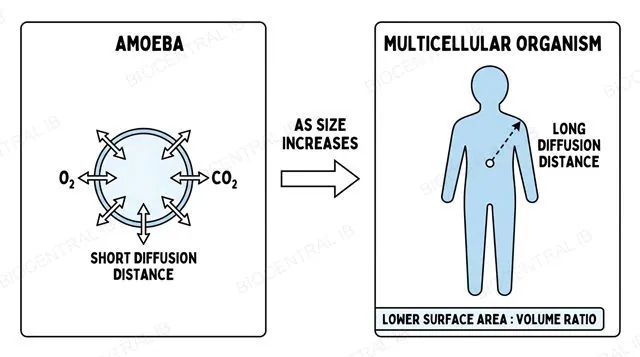
A small unicellular organism like Amoeba exchanging gas directly across its cell membrane, next to a much larger multicellular organism where the diffusion distance from core to surface is far greater, labeled with surface area to volume ratio
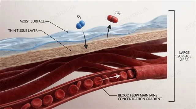
A generic gas exchange surface diagram: a thin moist membrane with gas molecules diffusing across a large surface area down a steep concentration gradient, labeled with permeability, thin tissue layer, moisture and large surface area
B3.1.2

What every exchange surface needs

Fick's law ties diffusion rate to surface area and concentration gradient, divided by diffusion distance — every efficient gas-exchange surface is built around maximising the first two and minimising the third.
  • A large surface area gives more sites for diffusion to happen at once; a thin, permeable tissue layer keeps the diffusion path as short as possible.
  • The surface must stay moist, since gases have to dissolve into a liquid film before crossing into cells, and a steep concentration gradient — kept up by blood flow and ventilation — keeps diffusion running fast rather than stalling near equilibrium.
Why it mattersLungs, gills and leaves look completely different, but every one of them is solving Fick's law the same way.
B3.1.3

Keeping the gradient steep

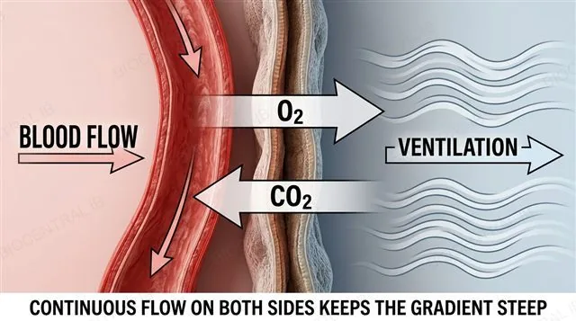
A concentration gradient only drives diffusion while it exists — animals maintain it at exchange surfaces with continuous blood flow and continuous ventilation.
  • A dense network of blood vessels constantly carries freshly oxygenated blood away and delivers CO₂-rich blood back to the surface, so the internal side of the gradient never runs out.
  • Ventilation — moving air through lungs or water through gills — constantly replaces the external side, keeping O₂ high and CO₂ low right where exchange happens.
Why it mattersWithout both sides moving, diffusion would slow to a crawl as the gradient flattened toward equilibrium.
A thin exchange membrane with blood flowing on one side via capillaries and ventilated air or water flowing on the other side, arrows showing O2 diffusing one way and CO2 the other, labeled with blood flow and ventilation maintaining the concentration gradient
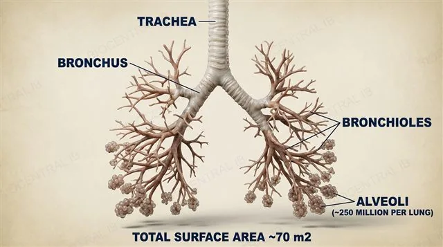
The branching airway of a mammalian lung from trachea to bronchi to bronchioles ending in clusters of alveoli, labeled
B3.1.4

From windpipe to 250 million alveoli

Air travels through a branching airway that turns one windpipe into an enormous number of tiny sacs, each one a gas-exchange surface.
  • Air passes from the trachea (held open by cartilage rings) into two bronchi, then repeatedly branches into progressively narrower bronchioles, whose smooth muscle can dilate or constrict to control airflow.
  • The bronchioles terminate in roughly 250 million alveoli per lung — together giving a total gas-exchange surface area of around 70 m², far more than could fit as a single smooth sac.
Why it mattersBranching is how the lung solves the surface-area problem: many tiny sacs beat one big one.
B3.1.4

The alveolus wall itself

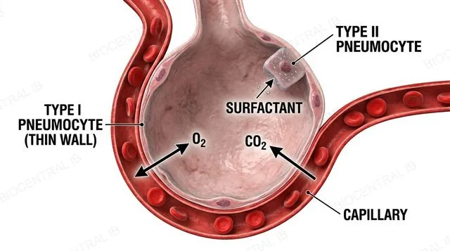
Each alveolus is wrapped in a dense capillary network and walled by a single layer of type I pneumocytes, with type II pneumocytes secreting surfactant into its lining.
  • Type I pneumocytes form an extremely thin single-cell wall, minimising the diffusion distance between air and blood; O₂ diffuses in from the alveolar air space while CO₂ diffuses out, each gas moving down its own gradient.
  • Type II pneumocytes secrete pulmonary surfactant, a phospholipid mixture that lowers surface tension in the moist alveolar lining and stops the alveolus collapsing on itself, especially during exhalation.
Why it mattersThin walls speed diffusion; surfactant keeps the sac from collapsing under its own surface tension — the alveolus needs both.
A single alveolus cross-section wrapped in capillaries, with a thin type I pneumocyte wall, a type II pneumocyte secreting surfactant, and arrows showing O2 diffusing into the capillary and CO2 diffusing out, labeled
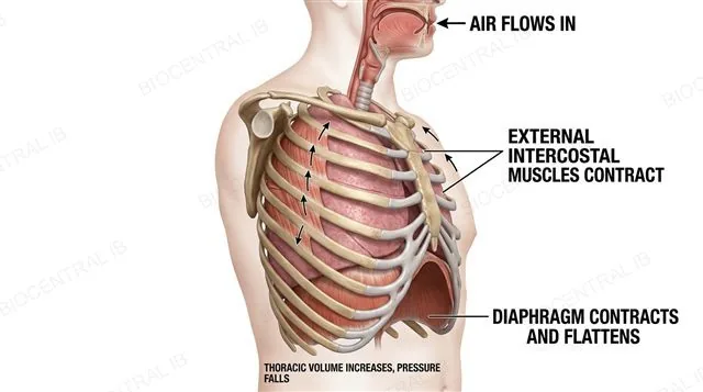
A cross section of the thorax during inspiration with the diaphragm contracted and flattened, the ribcage lifted up and out by contracted external intercostal muscles, and an arrow showing air flowing into the lungs, labeled
B3.1.5

Breathing in: expanding the thorax

Inspiration is an active process: the diaphragm and external intercostal muscles contract together to enlarge the thoracic cavity.
  • The diaphragm contracts and flattens downward while the external intercostal muscles contract, lifting the ribcage up and outward — together these increase the volume of the thoracic cavity.
  • As thoracic volume rises, pressure inside the lungs drops below atmospheric pressure, and air flows in down that pressure gradient until the two equalise.
Why it mattersEvery inhalation is Boyle's law in action: expand the container, and pressure inside falls below the pressure outside.
B3.1.5

Breathing out: passive or forced

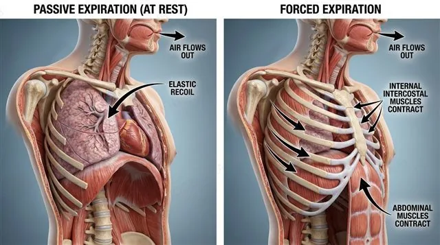
Expiration at rest is passive, but forced expiration recruits an entirely separate set of muscles — and the whole cycle is paced by the medulla oblongata.
  • At rest, the diaphragm and external intercostals simply relax; elastic recoil of the lungs and chest wall shrinks thoracic volume, raising pressure and pushing air back out — no muscle contraction needed.
  • Forced expiration (blowing out a candle) additionally recruits internal intercostal muscles, which pull the ribs down and in, and abdominal muscles, which push the diaphragm upward; breathing rate itself is set by the medulla oblongata responding to rising CO₂ and falling blood pH.
Why it mattersPassive expiration costs no energy — the lung is basically an elastic balloon that wants to deflate on its own.
A cross section of the thorax during forced expiration with internal intercostal muscles pulling the ribs down and abdominal muscles pushing the diaphragm upward, contrasted with a smaller passive expiration arrow from elastic recoil, labeled
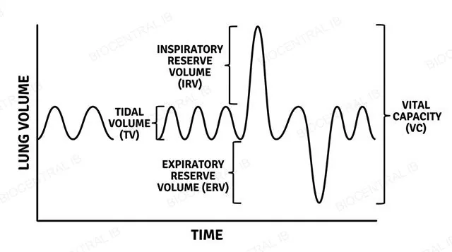
A spirometer trace (kymograph) over time showing tidal volume as small regular oscillations, with inspiratory reserve volume above and expiratory reserve volume below, and vital capacity spanning all three, labeled
B3.1.6

Measuring what the lungs can do

A spirometer traces the volume of air moved with each breath, letting four standard lung volumes be read directly off the graph.
  • Tidal volume (TV) is the air moved in one normal resting breath, about 500 mL; inspiratory and expiratory reserve volumes (IRV, ERV) are the extra air that can still be inhaled or exhaled beyond that.
  • Vital capacity (VC = TV + IRV + ERV) is the maximum air moved in a single breath, typically 4.5–5 L in an adult; if soda lime absorbs the CO₂ exhaled, the trace's slow downward drift reveals the rate of O₂ consumption.
Why it mattersTV and VC get mixed up constantly — TV is one normal breath, VC is the biggest breath you can physically take.
B3.1.7

A leaf has the same problem

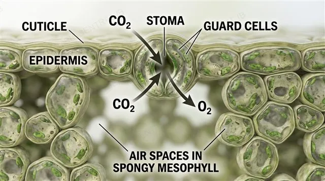
A leaf must let CO₂ in for photosynthesis while limiting water loss — and its structure is built around that trade-off.
  • A waxy cuticle over the epidermis blocks most gas and water movement, forcing exchange to happen through the stomata — pores whose aperture is controlled by a pair of guard cells around each one.
  • Once CO₂ is inside, large air spaces running through the spongy mesophyll let it diffuse quickly to every photosynthesising cell, while veins deliver water and carry sugars away.
Why it mattersA leaf and a lung solve the same physics with completely different materials — pores instead of alveoli, mesophyll air spaces instead of capillaries.
A leaf cross section showing a waxy cuticle over the epidermis, a stoma flanked by guard cells, CO2 diffusing in and O2 diffusing out through the stoma, and air spaces in the spongy mesophyll layer, labeled
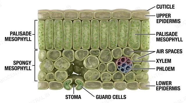
A full labeled transverse section of a dicotyledonous leaf showing cuticle, upper epidermis, palisade mesophyll, spongy mesophyll with air spaces, a vascular bundle with xylem and phloem, lower epidermis, and a stoma with guard cells, labeled
B3.1.8

A leaf, layer by layer

A transverse section of a dicotyledonous leaf shows five distinct tissue layers, each doing a different job.
  • The upper epidermis (covered by the cuticle) and lower epidermis (bearing most of the stomata) sandwich two mesophyll layers: tightly packed, chloroplast-rich palisade mesophyll just below the upper epidermis, and loosely arranged spongy mesophyll with large air spaces beneath it.
  • Vascular bundles run through the middle, with xylem toward the upper side delivering water and phloem toward the lower side carrying sugars away.
Why it mattersThis is a diagram IB expects you to be able to draw and label from memory, not just recognise.
B3.1.9

Transpiration: the price of open stomata

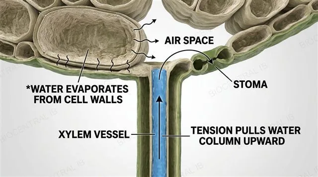
Transpiration — the evaporative loss of water vapour through open stomata — is an unavoidable side effect of keeping stomata open for CO₂ uptake.
  • A single stoma cannot let CO₂ diffuse in without also letting water vapour diffuse out, so any leaf built for gas exchange is automatically built to lose water; this loss also generates the tension that draws water up the xylem, cools the leaf by evaporation, and helps keep cells turgid.
  • Transpiration rate rises with higher temperature, higher light intensity, and moving air, and falls as humidity increases — all because these factors change how steep the water-vapour gradient is between the leaf's air spaces and the outside air.
Why it mattersTranspiration isn't a design flaw — it's the direct, physically necessary cost of the exact same stomata that let CO₂ in.
Water evaporating from mesophyll cell walls into a leaf's internal air spaces and escaping through an open stoma, with an arrow showing tension pulling a continuous water column up through the xylem toward the leaf, labeled
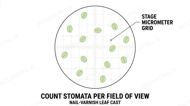
A microscope field of view showing a leaf epidermis nail-varnish cast with visible stomata being counted against a calibrated stage micrometer grid, labeled
B3.1.10

Counting stomata

Stomatal density — stomata counted per unit leaf area — can be measured directly from a leaf surface cast and compared across habitats.
  • Painting clear nail varnish onto the leaf surface, letting it dry, and peeling it off produces a cast of the epidermis that can be examined and counted under a microscope in a field of view calibrated with a stage micrometer.
  • Species from dry habitats typically evolve lower stomatal density than species from wetter habitats, since fewer stomata mean less water lost overall; repeating counts across several fields of view is what makes the result reliable, since real leaf tissue is naturally variable.
Why it mattersOne count from one field of view tells you almost nothing — the NOS point here is that biological variability demands replicate trials.
Quick check

A plant leaf loses a large amount of water through transpiration even when it doesn't need to cool down. Why can't the leaf just keep its stomata permanently closed to stop this water loss?

Because guard cells cannot physically stay closed for long periods
Because closed stomata would also block the CO₂ the leaf needs for photosynthesis
Because transpiration is required to open the stomata in the first place
Because the waxy cuticle cannot function without open stomata
Correct answer: closed stomata would also block CO₂ entry. The same pore that lets water vapour escape is the only route CO₂ has to enter for photosynthesis — a leaf cannot allow one gas through without allowing the other, so transpiration is the unavoidable cost of staying able to photosynthesise.
Part two · HL

Haemoglobin fine-tunes the exchange

B3.1.11 – B3.1.13
B3.1.11 · HL

Foetal haemoglobin out-competes maternal Hb

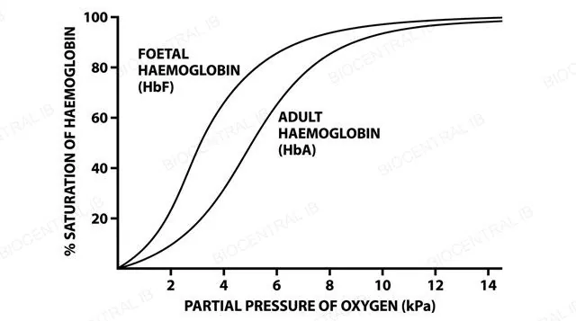
Foetal haemoglobin (HbF) has a higher affinity for O₂ than adult haemoglobin (HbA), letting a foetus pull oxygen across the placenta from its own mother's blood.
  • HbA is built from two α-globin and two β-globin chains; HbF instead pairs two α-globin chains with two γ-globin chains, a structural difference that gives HbF a higher O₂ affinity than HbA.
  • Because HbF's oxygen dissociation curve sits to the left of HbA's, at the same, relatively low placental pO₂, HbF binds O₂ more readily — producing a net transfer of O₂ from maternal blood into foetal blood across the placental exchange surface.
Why it mattersA higher-affinity molecule always wins a tug-of-war for O₂ at the same partial pressure — that's the entire mechanism of placental gas exchange.
Two oxygen dissociation curves on the same axes, percent saturation of haemoglobin against partial pressure of oxygen in kilopascals, with the foetal haemoglobin curve shifted to the left of the adult haemoglobin curve, labeled per the IB data booklet convention
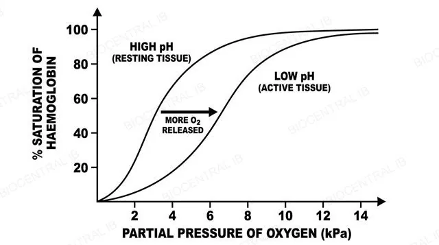
Two oxygen dissociation curves on the same axes, one for resting tissue at normal pH shifted left and one for actively respiring tissue at lower pH shifted right, with an arrow showing more oxygen released at the same partial pressure in the right-shifted curve, labeled
B3.1.12 · HL

The Bohr shift: CO₂ unlocks oxygen

The Bohr shift is the drop in haemoglobin's O₂ affinity caused by rising CO₂ and falling pH — and it happens exactly where the body needs O₂ released.
  • In actively respiring tissue, CO₂ dissolves into plasma and reacts with water to form carbonic acid, which dissociates into H⁺ and bicarbonate, lowering local pH.
  • That drop in pH (and the CO₂ itself) reduces haemoglobin's affinity for O₂, shifting its oxygen dissociation curve to the right, so more O₂ is released at the same pO₂ exactly in the tissues generating the most CO₂.
Why it mattersThe Bohr shift automatically routes more oxygen to whichever tissue is working hardest, with no nervous signal required — it's built into the haemoglobin molecule itself.
B3.1.13 · HL

Why the curve is S-shaped

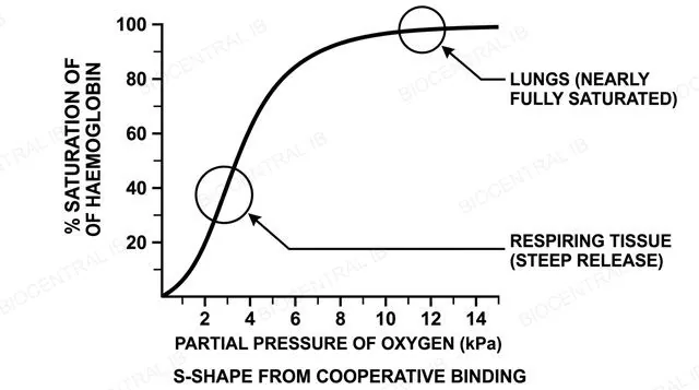
The oxygen dissociation curve's sigmoidal shape comes directly from cooperative binding between haemoglobin's four O₂-binding sites.
  • Binding the first O₂ molecule changes haemoglobin's conformation in a way that makes the next O₂ molecule easier to bind, and so on — this positive cooperativity produces the curve's steep middle section rather than a smooth, gradual rise.
  • In the lungs, where pO₂ is high (around 12–13 kPa), the curve sits on its flat plateau and haemoglobin is nearly fully saturated; in respiring tissue, where pO₂ is low (around 2–3 kPa), the curve is at its steepest, so a small drop in pO₂ releases a large amount of O₂.
Why it mattersThe steep middle section isn't a coincidence — it's positioned exactly where tissues sit on the pO₂ scale, so haemoglobin unloads O₂ efficiently right where it's needed.
A single S-shaped sigmoidal oxygen dissociation curve, percent saturation of haemoglobin on the y axis against partial pressure of oxygen in kilopascals on the x axis, with the flat plateau region near 12 to 13 kPa labeled lungs and the steep middle region near 2 to 3 kPa labeled respiring tissue, labeled
Quick check · HL

Blood from resting muscle and blood from the same muscle during hard exercise are both exposed to the same partial pressure of oxygen. Which one has haemoglobin with the lower O₂ affinity, and why?

Resting muscle blood — because resting tissue has more O₂ already bound
Exercising muscle blood — higher CO₂ and lower pH there shift the dissociation curve right, cutting O₂ affinity
They are identical — O₂ affinity does not change with CO₂
Exercising muscle blood — because muscle contraction directly binds to haemoglobin
Correct answer: exercising muscle blood has the lower affinity. Hard-working muscle produces more CO₂, which lowers local pH and shifts haemoglobin's oxygen dissociation curve to the right — the Bohr shift — reducing its affinity for O₂ so more is released exactly where respiration is fastest.

Key vocabulary

Worth being able to define in a single sentence each

Gas exchange
Diffusion of O₂ into and CO₂ out of an organism, driven by a concentration gradient.
Ventilation
Physical movement of air or water over a gas-exchange surface to maintain a gradient.
Vital capacity
The maximum volume of air moved in one breath: TV + IRV + ERV.
Guard cells
Paired cells controlling a stoma's aperture by active K⁺ transport and osmosis.
Transpiration
Evaporative loss of water vapour from a plant, mainly through open stomata.
Bohr shift HL
Rightward shift of the O₂ dissociation curve caused by rising CO₂ and falling pH.
Oxygen dissociation curve HL
Graph of % haemoglobin saturation against pO₂, S-shaped from cooperative binding.

Where this shows up again

B1.1
The alveoli are lined with a thin film of water containing surfactant (a phospholipid mixture). How do the properties of phospholipids (B1.1) enable surfactant to reduce surface tension at the air-water interface?
B2.1
Oxygen diffuses from the alveoli into the blood, and CO₂ diffuses in the opposite direction. How does the structure of the cell membrane (B2.1) facilitate the rapid diffusion of these small, nonpolar gas molecules?
B3.2
The circulatory system (B3.2) transports oxygen from the lungs to respiring tissues. Explain how the Bohr effect ensures that oxygen is released preferentially in tissues with the highest metabolic rate.
C1.2
During aerobic respiration (C1.2), cells consume O₂ and produce CO₂. Explain how the ventilation rate is homeostatically regulated to match O₂ delivery and CO₂ removal to the rate of cellular respiration.

B3.1 Gas exchange — one-page recap

Screenshot this slide to revise from

The core problem
  • SA:V falls as organisms get bigger.
  • Fick's law: area × gradient ÷ distance.
Mammalian lung
  • Trachea → bronchi → bronchioles → ~250M alveoli.
  • Type I thin wall; type II secretes surfactant.
Ventilation & volumes
  • Inspiration active; rest expiration passive.
  • Spirometer: TV, IRV, ERV, VC.
Leaf structure
  • Cuticle → epidermis → palisade → spongy → vein.
  • Stomata + guard cells control gas flow.
Transpiration & density
  • Water loss is the unavoidable cost of CO₂ uptake.
  • Nail-varnish cast + stage micrometer counts stomata.
Haemoglobin HL
  • HbF left-shifted vs HbA — placental transfer.
  • Bohr shift: ↑CO₂/↓pH shifts curve right.

Every surface, the same physics

A lung, a leaf and a single red blood cell are all fighting the same battle — more area, shorter distance, steeper gradient.
B3.1 Gas exchange · BioCentral IB
Use ↓ ↑ or click to navigate
01 / 22