BioCentral IBBioCentral IB
IB BIOLOGY · THEME B · B3.2

Transport

A giraffe's heart has to generate enough pressure to push blood two metres uphill to its brain, while a tree pulls water past the same height with no pump at all — animals and plants solve the same problem, fluid transport, with completely different physics.
Guiding questions

What adaptations facilitate transport of fluids in animals and plants?

What are the differences and similarities between transport in animals and plants?

Part one

Moving blood, moving sap

B3.2.1 – B3.2.10
B3.2.1

The capillary: exchange at its narrowest

Capillaries are the site of nearly all material exchange between blood and tissue — a highly branched network of the narrowest, thinnest vessels in the body.
  • Their wall is a single layer of endothelial cells, minimising diffusion distance, and the lumen is barely wider than a red blood cell — forcing cells through in single file and maximising the time each one spends in contact with the wall.
  • Extensive branching gives the whole capillary bed an enormous total surface area and slows overall flow rate; small pores between endothelial cells — and, in some beds, larger fenestrations — let fluid and small solutes cross rapidly exactly where exchange must be fastest.
Why it mattersEvery adaptation here is Fick's law again: shrink the diffusion distance, grow the exchange area.
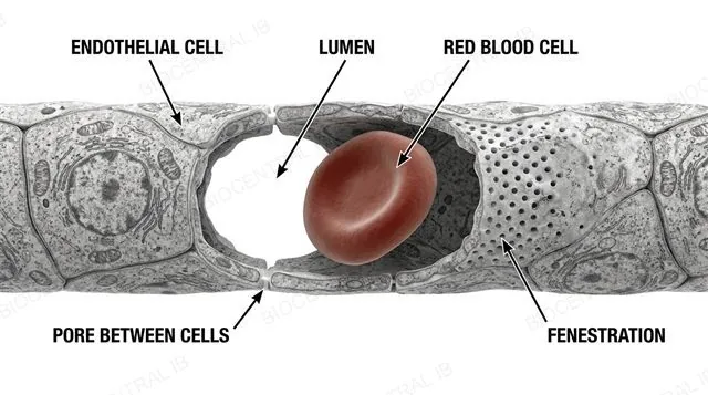
A capillary cross-section with a single layer of endothelial cells, a narrow lumen with a red blood cell passing through in single file, small pores between adjacent endothelial cells, and a fenestration in one region, labeled
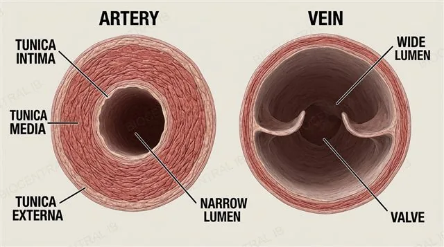
Side-by-side cross sections of an artery and a vein, each showing tunica intima, tunica media and tunica externa layers, with the artery having a thick tunica media and narrow lumen and the vein having a thin tunica media, wide lumen and an internal valve, labeled
B3.2.2

Same three layers, different proportions

Arteries and veins are both built from the same three-layer wall plan, but the thickness of each layer — and the width of the lumen — reveal which vessel is which under a microscope.
  • Every blood vessel shares a tunica intima (inner endothelium), tunica media (smooth muscle and elastic fibres) and tunica externa (outer connective tissue) — what differs between vessel types is proportion, not which layers are present.
  • In a micrograph, an artery shows a thick tunica media relative to a narrow lumen, while a vein shows a thin tunica media around a wide, often flattened lumen — this wall-to-lumen ratio, not overall vessel size, is how the two are told apart.
Why it mattersIB expects you to identify an unlabelled vessel from a micrograph using exactly this ratio — not from size or colour.
B3.2.3

Arteries: built to survive the surge

Arteries carry blood away from the heart under high, pulsatile pressure, and their wall is adapted to withstand and smooth that surge.
  • Abundant smooth muscle in the tunica media allows vasoconstriction and vasodilation, adjusting vessel diameter and resistance and helping regulate blood pressure.
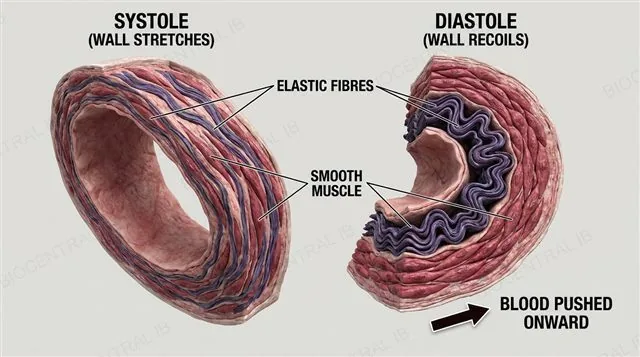
  • Elastic fibres stretch as each heartbeat forces blood in (systole) and recoil once the heart relaxes (diastole), stopping pressure from collapsing to zero between beats and converting a pulsing surge into a steadier flow downstream.
Why it mattersElastic recoil is why blood pressure never drops to zero between heartbeats — the artery wall itself keeps pushing.
An artery wall cross section showing thick smooth muscle and elastic fibres in the tunica media stretching during systole and recoiling during diastole, with an arrow showing blood being pushed onward by the elastic recoil, labeled
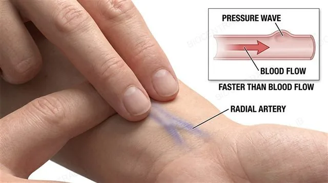
A hand with two fingertips palpating the radial artery pulse at the wrist, next to a diagram of a pressure wave travelling rapidly along an artery wall ahead of the slower-moving blood itself, labeled
B3.2.4

The pulse is a pressure wave, not blood itself

A pulse is the pressure wave generated by each contraction of the left ventricle, travelling through the elastic arterial wall faster than the blood inside it actually moves.
  • It can be felt wherever an artery runs close to the skin over a firm surface such as bone — most commonly the radial artery at the wrist or the carotid artery in the neck.
  • Counting beats over a fixed time gives heart rate in beats per minute, whether measured traditionally by fingertip palpation or digitally with a pulse sensor — the two methods can be directly compared for accuracy and consistency.
Why it mattersA pulse reading is really a heart-rate measurement in disguise — it works because the pressure wave, not the blood, reaches your fingertip almost instantly.
B3.2.5

Veins: built to return blood uphill

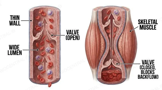
Veins carry blood back to the heart at low pressure, so their structure trades wall strength for a wide, low-resistance channel and one-way valves.
  • A thin tunica media with little smooth muscle or elastic tissue reflects the low internal pressure; a wide lumen minimises resistance to the sluggish flow.
  • Internal pocket valves prevent backflow, while contraction of surrounding skeletal muscle compresses the vein and pushes blood toward the heart — the skeletal muscle pump — and pressure changes in the thorax during breathing add a further push, the respiratory pump.
Why it mattersWithout valves, gravity alone would pull blood in your legs back downward every time those leg muscles relaxed.
A vein cross section with a thin wall, wide lumen, and an internal pocket valve, next to a diagram of a contracting skeletal muscle compressing the vein and pushing blood toward the heart while the valve prevents backflow, labeled
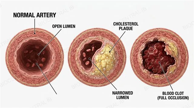
A coronary artery cross section progressing through three stages from a normal open lumen, to a cholesterol plaque narrowing the lumen, to a ruptured plaque with a blood clot fully occluding the vessel, labeled
B3.2.6

When a coronary artery closes

Atherosclerosis is the build-up of cholesterol-rich plaques inside an artery wall, and in a coronary artery it can escalate all the way to a heart attack.
  • Plaques narrow the lumen and stiffen the wall over years, driven by risk factors including high LDL cholesterol, smoking, hypertension, diabetes, poor diet and a sedentary lifestyle.
  • If a plaque ruptures, a blood clot can form and fully occlude the artery — cutting off oxygen to part of the heart muscle and causing myocardial infarction, with localised cell death (necrosis).
Why it mattersThe coronary arteries are the heart's own blood supply — block them, and the pump keeping every other organ alive starts failing itself.
B3.2.6 · NOS

A strong correlation still isn't proof

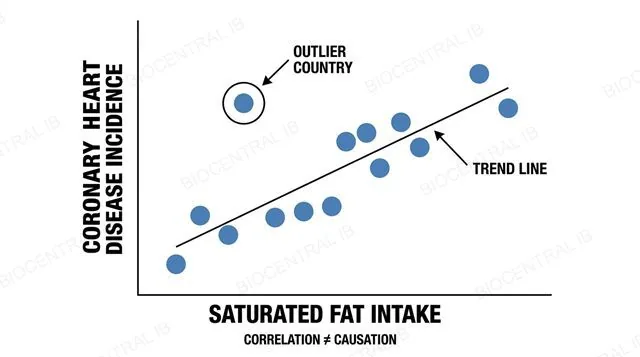
Correlation coefficients quantify how strongly two variables move together, but even a strong correlation between two variables does not, by itself, prove that one causes the other.
  • Epidemiological data show a strong correlation between saturated fat intake and coronary heart disease incidence across populations — consistent with a causal link, but not sufficient to establish one on its own.
  • A low or near-zero correlation coefficient can be strong evidence against a hypothesis; but confirming causation instead requires more — a plausible biological mechanism, controlled studies, and ruling out confounding variables.
Why it mattersThis exact example — fat intake and heart disease — is IB's standard case for "correlation isn't causation": it can weaken a claim fast, but it can never finish proving one.
A scatter graph plotting average saturated fat intake against coronary heart disease incidence for several countries, with a positive trend line and a correlation coefficient labeled, and one outlier country marked to show that correlation alone is not proof of causation, labeled
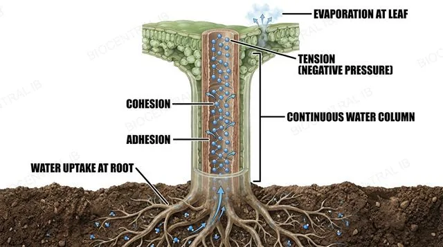
A cross section of a plant from root through stem to leaf showing a continuous water column inside the xylem, tension generated at the leaf by evaporation from mesophyll cell walls, cohesion between water molecules shown as hydrogen bonds holding the column together, and adhesion of water to the xylem walls, labeled
B3.2.7

Cohesion-tension: pulling water without a pump

A continuous column of water is pulled from root to leaf entirely by transpiration — evaporation at the top generates tension that cohesion transmits all the way down.
  • Water evaporating from mesophyll cell walls draws more water out of the xylem and through the cell wall by capillary action, generating tension — a negative pressure potential — at the top of the column.
  • Hydrogen bonds between water molecules (cohesion) hold the column together under that tension so it doesn't break, while adhesion of water to the hydrophilic xylem walls helps resist gravity's pull as the whole column rises.
Why it mattersNo cell anywhere in the plant is actively pumping this water — the entire lift comes from physics happening at the leaf's surface.
B3.2.8

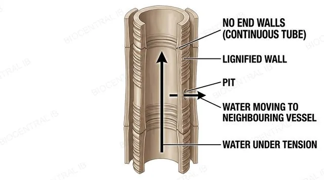
The xylem vessel: dead tubes built to not break

A mature xylem vessel is a dead, hollow tube whose structure exists entirely to move water under tension without collapsing or leaking.
  • At maturity, xylem vessel elements have no cytoplasm and incomplete or absent end walls, joining end-to-end into a continuous, unobstructed tube that offers minimal resistance to flow.
  • Lignified walls give the rigidity needed to withstand the strong tension of transpiration pull without collapsing inward, while pits — thin, unlignified regions in the wall — allow water to move laterally into neighbouring vessels and surrounding cells.
Why it mattersA living cell couldn't survive what a xylem vessel experiences — which is exactly why the vessel gives up being alive to do the job.
A xylem vessel formed from stacked dead vessel elements with no end walls, thick lignified walls, and a pit allowing lateral water movement into a neighbouring vessel, with an arrow showing water under tension moving upward, labeled
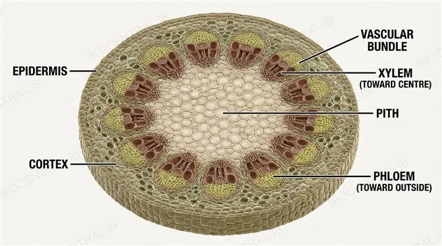
A transverse section of a dicotyledonous stem showing an outer epidermis, a cortex, a ring of vascular bundles each with xylem toward the centre and phloem toward the outside, and a central pith, labeled
B3.2.9

A dicot stem, ring by ring

A transverse section of a dicotyledonous stem shows vascular bundles arranged in a ring, with each tissue positioned for what it delivers or protects.
  • An outer epidermis, often covered by a cuticle, protects the stem; a cortex of ground tissue lies between it and the ring of vascular bundles.
  • Each vascular bundle carries xylem toward the stem's centre and phloem toward its outside, with a central pith of parenchyma providing storage — a diagram IB expects you to draw and label directly from a micrograph.
Why it mattersXylem-in, phloem-out is the stem's signature arrangement — get the ring the wrong way round and every downstream inference about flow direction breaks.
B3.2.10

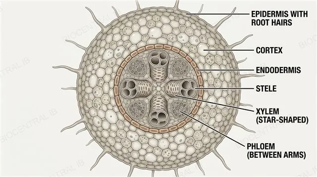
A dicot root: the star at the centre

A transverse section of a dicotyledonous root looks nothing like the stem — its vascular tissue sits in a central star-shaped stele, not a peripheral ring.
  • An epidermis bearing root hairs, which extend surface area for water and ion uptake, surrounds a cortex, with an endodermis forming the innermost cortical layer.
  • The central stele holds xylem arranged in a star or cross shape with phloem in separate patches between its arms — a layout that can be drawn directly from a micrograph and annotated with each tissue's function.
Why it mattersThe root's central xylem star versus the stem's peripheral ring is one of the most reliable ways IB tests whether you can actually read a plant micrograph, not just recall a labelled textbook diagram.
A transverse section of a dicotyledonous root showing an epidermis with root hairs, a cortex, an endodermis, and a central stele with xylem arranged in a star shape and phloem in patches between the arms, labeled
Quick check

A micrograph shows a blood vessel with a thin wall relative to a wide lumen, and a visible internal valve. Which vessel is this, and why doesn't it need a thick, muscular wall?

An artery — arteries always have thin walls relative to their lumen
A vein — it carries blood at low pressure, so it doesn't need to resist a high-pressure surge
A capillary — capillaries have valves to control single-file blood flow
An artery — the valve regulates vasoconstriction
Correct answer: a vein. Blood returning to the heart is at low, steady pressure, so a vein's wall needs far less smooth muscle and elastic tissue than an artery's — the wide lumen minimises resistance instead, and the valve (something arteries and capillaries don't have) stops that low-pressure flow running backward.
Part two · HL

Fluid balance, circulation and the heartbeat

B3.2.11 – B3.2.18
B3.2.11 · HL

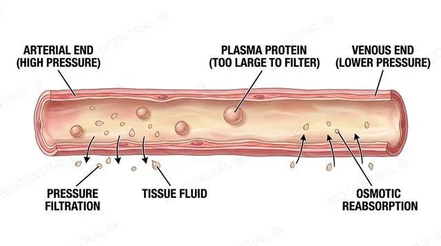
Tissue fluid: leaking out, soaking back in

Tissue fluid forms and is reabsorbed at opposite ends of the same capillary, driven by a hydrostatic pressure that falls along its length.
  • At the arterial end, high hydrostatic pressure — generated by ventricular contraction — forces plasma fluid out through the capillary's pores by pressure filtration, forming tissue fluid; large plasma proteins such as albumin are too big to follow and stay behind in the blood.
  • By the venous end, hydrostatic pressure has dropped, and the osmotic (oncotic) pressure created by those retained proteins now draws water back into the capillary — reabsorption; roughly 90% of the fluid filtered out returns this way.
Why it mattersThe same capillary filters fluid out at one end and pulls most of it back in at the other, purely because pressure changes along its length.
A single capillary running from its arterial end to its venous end, with an arrow showing high hydrostatic pressure forcing plasma fluid out through pores near the arterial end and an arrow showing osmotic pressure from retained plasma proteins drawing water back in near the venous end, labeled
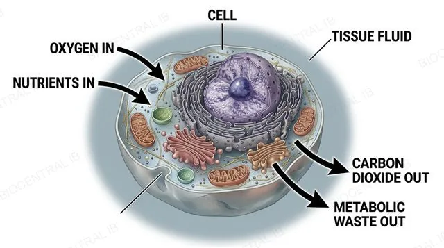
A tissue cell surrounded by tissue fluid, with arrows showing oxygen and nutrients diffusing from the tissue fluid into the cell and carbon dioxide and metabolic waste diffusing from the cell into the tissue fluid, labeled
B3.2.12 · HL

Tissue fluid: the middleman for every cell

Tissue fluid is plasma minus its large proteins and cells, and it is this fluid — not blood itself — that every body cell actually exchanges materials with.
  • Oxygen and nutrients such as glucose and amino acids diffuse from tissue fluid into cells, moving down a concentration gradient the cells themselves maintain by continuously consuming these substances.
  • Carbon dioxide and other metabolic wastes diffuse the opposite way, from cells into tissue fluid, because respiration keeps their intracellular concentration higher — no active transport is needed across the capillary wall itself.
Why it mattersNo body cell ever actually touches blood — tissue fluid is the only interface a cell has with the entire circulatory system.
B3.2.13 · HL

Where the leftover 10% goes

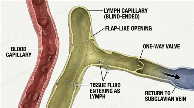
The tissue fluid that isn't reabsorbed at the capillary's venous end doesn't just accumulate — it drains into a separate one-way vessel network.
  • Excess tissue fluid enters blind-ended lymph capillaries through flap-like openings in their thin walls, and is now called lymph rather than tissue fluid.
  • Lymph vessels carry one-way valves and are propelled mainly by surrounding skeletal muscle contraction; lymph passes through lymph nodes for immune surveillance before draining back into the bloodstream at the subclavian veins.
Why it mattersWithout this drainage route, that unreabsorbed 10% would accumulate in tissues every single day — the lymphatic system is what keeps fluid balance from failing.
A blind-ended lymph capillary next to a blood capillary, with excess tissue fluid entering the lymph capillary through flap-like openings, a one-way valve in the lymph vessel, and an arrow showing lymph draining toward a subclavian vein, labeled
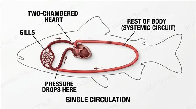
A simple circuit diagram of a bony fish's single circulation, showing a two-chambered heart pumping blood through the gills where pressure drops and then on to the rest of the body before returning to the heart, labeled
B3.2.14 · HL

A fish: one circuit, one pressure drop

A bony fish has a single circulation: blood passes through the heart just once per full circuit of the body.
  • A two-chambered heart pumps blood through the gills, where the resistance of the narrow gill capillaries causes a substantial drop in pressure as gases are exchanged.
  • That already-reduced pressure is all that's left to drive blood around the rest of the systemic circuit before it returns to the heart — a hard ceiling on how much pressure, and therefore how fast a metabolic rate, the system can support.
Why it mattersLosing pressure at the gills isn't a design flaw — it's the direct cost of having only one heart pump to serve two circuits in series.
B3.2.14 · HL

A mammal: two circuits, re-pressurised

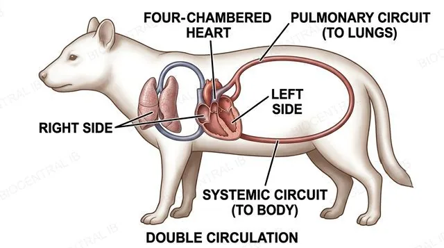
A mammal's double circulation separates gas exchange from systemic delivery by sending blood through the heart twice per full circuit.
  • A four-chambered heart keeps a pulmonary circuit (right side, to the lungs) and a systemic circuit (left side, to the body) completely separate.
  • Blood returning from the lungs is re-pressurised by the left ventricle before it is sent to the body, so pressure lost at the gas-exchange surface never limits pressure in the systemic circuit — supporting the higher metabolic rate that endothermy demands.
Why it mattersDecoupling the two circuits is the single structural change that lets a mammal run its whole body at far higher pressure than any fish could.
A simple circuit diagram of a mammal's double circulation, showing a four-chambered heart with a separate pulmonary circuit to the lungs on the right side and a separate systemic circuit to the body on the left side, with blood re-pressurised by the left ventricle after returning from the lungs, labeled
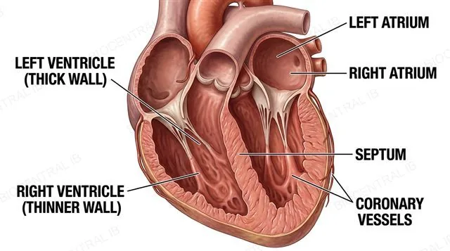
A frontal-plane cross section of the mammalian heart showing the four chambers, a muscular septum separating the left and right sides, a much thicker left ventricle wall than right ventricle wall, and coronary vessels branching from the base of the aorta over the heart's own surface, labeled
B3.2.15 · HL

The heart's own walls, unequal by design

The mammalian heart's chamber walls, septum and own blood supply are all shaped by the different pressures each side must generate.
  • The left ventricle's wall is far thicker than the right ventricle's, since it must generate enough pressure to drive blood around the entire systemic circuit, while the right side only pressurises blood as far as the nearby lungs; a muscular septum keeps oxygenated and deoxygenated blood from mixing between the two sides.
  • Coronary arteries branch from the base of the aorta and run over the heart's own surface, supplying the hard-working, densely mitochondria-packed cardiac muscle with the oxygenated blood it needs to keep contracting continuously.
Why it mattersThe heart is the one organ that has to supply itself — if its own coronary vessels fail, the pump that keeps every other tissue alive stops first.
B3.2.15 · HL

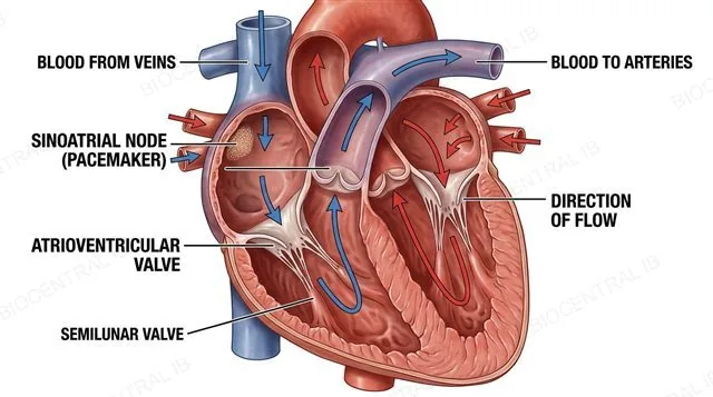
Valves and a pacemaker: flow that only goes one way

Two sets of one-way valves and a self-generating pacemaker together make sure the heart's contraction is both unidirectional and correctly timed.
  • Atrioventricular valves (tricuspid, bicuspid) sit between atria and ventricles, and semilunar valves (aortic, pulmonary) sit between ventricles and their arteries; both sets open and close purely in response to pressure differences on either side, never actively pumping themselves.
  • The sinoatrial node, a patch of specialised cardiac muscle in the right atrium wall, generates its own rhythmic electrical impulses — the heartbeat's pacemaker — initiating and coordinating the contraction sequence traced from named veins to named arteries.
Why it mattersA valve that only responds to pressure needs no nerve signal to work correctly — the heart's own internal plumbing enforces one-way flow automatically.
A frontal-plane diagram of the mammalian heart tracing the unidirectional flow of blood from named veins through the atrioventricular valves into the ventricles and out through the semilunar valves into named arteries, with the sinoatrial node marked in the wall of the right atrium as the pacemaker, labeled
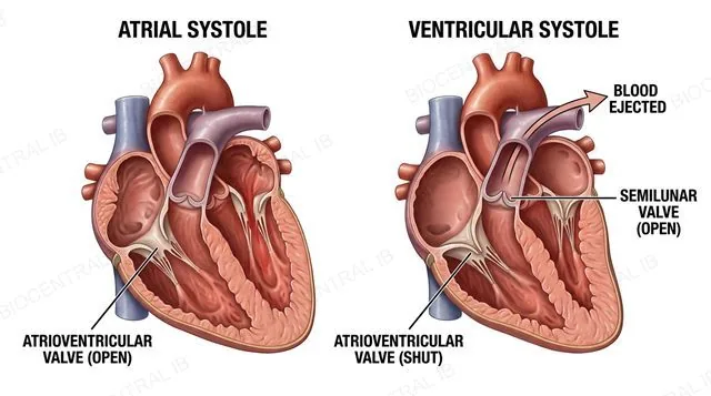
A sequence diagram of the left side of the heart during atrial systole, showing the atrium contracting with open atrioventricular valves and closed semilunar valves, followed by ventricular systole showing the ventricle contracting with the atrioventricular valves forced shut and the semilunar valves pushed open as blood is ejected into the aorta, labeled
B3.2.16 · HL

Systole: atria first, then ventricles

The cardiac cycle's contraction phase happens in two sequential stages, each with its own valve behaviour.
  • Atrial systole begins after the ventricles are already around 70% full from passive filling; the atria contract to push in the final volume of blood, with the AV valves open and the semilunar valves still closed.
  • Ventricular systole follows: rising ventricular pressure forces the AV valves shut, producing the first heart sound ("lub"), and once that pressure exceeds the pressure in the arterial trunk, the semilunar valves are pushed open and blood is ejected.
Why it mattersEvery valve in this sequence opens and closes purely because of the pressure difference across it — nothing here is directly triggered by a nerve signal.
B3.2.16 · HL

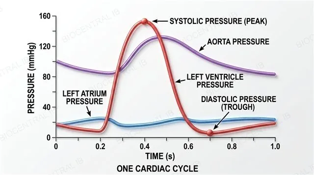
Diastole, and reading the pressure trace

Diastole resets the cycle, and the pressure changes across the whole sequence are exactly what a blood pressure reading measures.
  • In diastole, the ventricles relax; once ventricular pressure drops below arterial pressure, the semilunar valves close (the second heart sound, "dub"), and once it falls further, below atrial pressure, the AV valves open and passive filling of the ventricles begins again.
  • A blood pressure reading (e.g. 120/80 mmHg) reports systolic pressure (the peak, during ventricular systole) over diastolic pressure (the trough, during diastole) — both values can be read directly off a pressure-time graph for the left ventricle and aorta.
Why it mattersSystolic and diastolic numbers are not two different measurements from two different moments picked at random — they are literally the highest and lowest points of the same continuous pressure trace.
A pressure-time graph over one cardiac cycle showing the left atrium, left ventricle and aorta pressure curves, with the ventricular pressure peak labeled systolic pressure and its trough during diastole labeled diastolic pressure, and the timing of semilunar and atrioventricular valve opening and closing marked, labeled
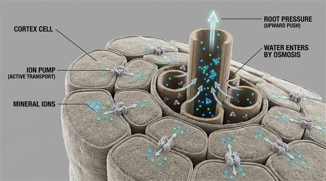
A root cross section showing cortex cells actively pumping mineral ions into the stele using membrane proton pumps, ions accumulating in the xylem, and water entering the xylem by osmosis, generating a positive root pressure pushing water upward, labeled
B3.2.17 · HL

Root pressure: a backup, not the main pump

Root pressure is a positive pressure generated by active ion transport in root cells, distinct from — and much weaker than — the tension driving transpiration pull.
  • Root cortex cells actively pump mineral ions such as K⁺ and NO₃⁻ into the stele using membrane transport proteins and ATP; as ions accumulate in the xylem, water follows by osmosis, generating a positive pressure that pushes water upward from below.
  • Root pressure only becomes the dominant driver of water movement when transpiration is negligible — for example at night, in high humidity, or in spring before deciduous leaves have opened; the rest of the time, transpiration pull (B3.2.7) does the real work.
Why it mattersRoot pressure being weak is the point — it's a fallback mechanism for when the plant's main pulling force from the leaves temporarily switches off.
B3.2.18 · HL

Sieve tube elements: stripped down for flow

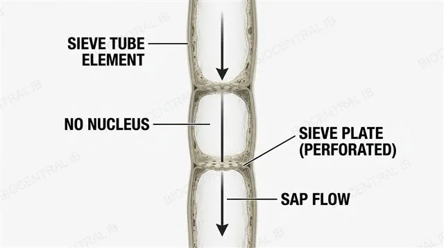
A sieve tube element is a living cell reduced to almost nothing but a channel, built to let sugary sap move through it with as little obstruction as possible.
  • It has no nucleus and greatly reduced cytoplasm and organelles, clearing internal space that would otherwise obstruct the mass flow of sap.
  • Sieve tube elements join end-to-end through sieve plates — perforated cross-walls — which still allow bulk flow of sap between adjacent elements while keeping the tube structurally continuous.
Why it mattersLosing a nucleus is a drastic step for a living cell to take — it only makes sense because this cell's entire job is to not get in the way of flow.
A row of sieve tube elements joined end to end through perforated sieve plates, each element shown with no nucleus and greatly reduced cytoplasm, with an arrow showing sap flowing through the sieve plate pores, labeled
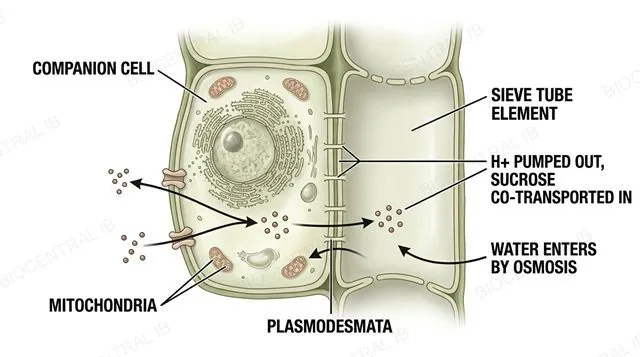
A companion cell connected to an adjacent sieve tube element by plasmodesmata, actively pumping hydrogen ions out and using the gradient to co-transport sucrose into the sieve tube at a source, with water following by osmosis to raise hydrostatic pressure, and a contrasting unloading region at a sink where sucrose and water leave and pressure falls, labeled
B3.2.18 · HL

Companion cells power the pressure-flow

Companion cells supply the active transport that sieve tube elements can no longer perform themselves, driving the pressure-flow mechanism of translocation.
  • A companion cell keeps its nucleus, dense cytoplasm and many mitochondria, and connects to its neighbouring sieve tube element through plasmodesmata — it does the metabolic work the sieve tube element gave up.
  • At a source, the companion cell pumps H⁺ out and uses the resulting gradient to co-transport sucrose into the sieve tube; the sucrose lowers water potential there, drawing water in by osmosis and raising hydrostatic pressure, which drives bulk flow of sap toward a sink, where sucrose and water both leave and pressure falls.
Why it mattersThe pressure difference between source and sink — not any pumping inside the tube itself — is the entire engine behind phloem transport.
Quick check · HL

A fish's blood pressure drops significantly after passing through the gills, before it reaches the rest of the body. Why doesn't a mammal have the same problem, despite also sending blood through the lungs before the body?

Mammals don't lose any pressure at all when blood passes through the lungs
The left ventricle re-pressurises blood after it returns from the lungs, in a separate systemic circuit
Mammal tissues simply need less blood pressure than fish tissues do
Fish gills generate extra pressure that fully compensates for what's lost
Correct answer: the left ventricle re-pressurises blood after the lungs. In a mammal's double circulation, blood returns to the heart after the (lower-pressure) pulmonary circuit and is boosted back up to high pressure by the left ventricle before starting the systemic circuit — decoupling gas-exchange pressure loss from systemic delivery pressure, something a fish's single circulation cannot do.

Key vocabulary

Worth being able to define in a single sentence each

Tunica media
Middle vessel-wall layer of smooth muscle and elastic fibres; its thickness differs sharply between artery and vein.
Pulse
The pressure wave from ventricular contraction, travelling through artery walls faster than the blood itself.
Atherosclerosis
Build-up of cholesterol-rich plaques in an artery wall, narrowing the lumen.
Cohesion-tension theory
Evaporation at the leaf generates tension; cohesion between water molecules transmits it down the xylem.
Casparian strip
Suberin band in the root endodermis that blocks the apoplast, forcing water through a cell membrane.
Double circulation HL
Separate pulmonary and systemic circuits, letting blood be re-pressurised after the lungs.
Pressure-flow hypothesis HL
Active loading of sucrose at a source raises pressure, driving bulk flow of phloem sap to a sink.

Where this shows up again

B3.1
The alveoli are wrapped in a dense capillary network (B3.1). How do the large surface area and thin walls of capillaries described here support the rapid gas exchange requirements of the alveolus?
B2.1
Root hair cells take up mineral ions using membrane transport proteins (B2.1). How does the structure of the cell membrane enable the active transport that generates root pressure?
C1.2
Cardiac muscle relies on continuous aerobic cell respiration (C1.2) to keep contracting without fatigue. Explain how the coronary circulation described here keeps the heart's own oxygen supply matched to its high respiratory demand.
C3.1
Heart rate is under feedback control from baroreceptors and chemoreceptors (C3.1). Explain how nervous control of heart rate could adjust the cardiac cycle described here to meet a sudden rise in the body's oxygen demand.

B3.2 Transport — one-page recap

Screenshot this slide to revise from

Blood vessels
  • Capillaries: thin, branched, some fenestrated.
  • Arteries: thick + elastic; veins: thin + valves.
Pulse & the coronary problem
  • Pulse = pressure wave, not blood flow.
  • Atherosclerosis → MI; correlation ≠ causation.
Xylem & plant tissues
  • Cohesion-tension pulls water; no pump needed.
  • Stem: xylem in, ring. Root: xylem star, centre.
Tissue fluid & lymph HL
  • Filtered out (arterial end), reabsorbed (venous end).
  • Excess drains via lymph back to the blood.
Circulation & the heart HL
  • Single (fish) vs double (mammal) circulation.
  • Valves + SA node pacemaker; systole then diastole.
Root pressure & phloem HL
  • Root pressure: weak backup, ion pumping.
  • Pressure-flow: sieve tubes + companion cells.

Every fluid, the same physics

A heart squeezing blood and a leaf evaporating water are doing the exact same thing — creating a pressure difference and letting physics move the fluid.
B3.2 Transport · BioCentral IB
Use ↓ ↑ or click to navigate
01 / 22