BioCentral IBBioCentral IB
IB BIOLOGY · THEME B · B3.3

Muscle and motility

A sarcomere only knows how to do one thing — pull. Everything from a heartbeat to a whale's dive is that single pulling motion, organised by skeletons, joints and nerves into the full range of animal movement.
Guiding questions

How do muscles contract and cause movement?

What are the benefits to animals of having muscle tissue?

Part one · HL

The sliding filament mechanism

B3.3.1 – B3.3.4
B3.3.1 · HL

Movement is a spectrum, not a switch

Movement — including internal movement, not just travel from place to place — occurs in essentially all living organisms, though not always at the speed the word "movement" suggests.
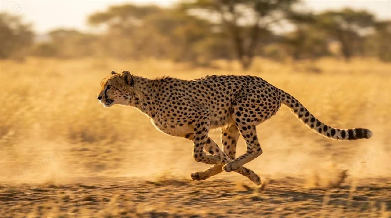
  • The brown-throated three-toed sloth is a fully motile mammal, capable of walking and climbing, but moves extremely slowly — an energy-saving adaptation suited to its low-calorie leaf diet, not a limitation on its ability to move.
  • Comparing a sloth's crawl to a sprinting mammal shows the same underlying capacity for movement expressed at wildly different speeds, each shaped by what that species actually needs to survive.
Why it mattersSpeed is a strategy, not a measure of whether an organism "really" moves.

A three-toed sloth climbing slowly through a tree beside a fast-running mammal, contrasting movement speed as a spectrum, photorealistic
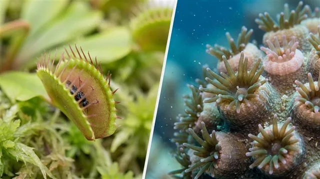
A Venus flytrap closing its trap on an insect and a coral polyp extending its tentacles, side by side, photorealistic
B3.3.1 · HL

Even "rooted" organisms move

Organisms often assumed to be immobile still show real, sometimes rapid, movement — the motile-versus-sessile divide is blurrier than it looks.
  • A Venus flytrap closes its trap within a fraction of a second when trigger hairs are touched, one of the fastest movements in the plant kingdom; corals extend and retract their polyps and tentacles to feed.
  • Most plants show slower tropisms — directional growth movements toward light (phototropism) or in response to gravity (gravitropism) — movement measured in hours or days rather than seconds.
Why it matters"Sessile" describes where an organism is rooted, not whether it can move — the two are not the same claim.
B3.3.2 · HL

Muscle is built in a strict hierarchy

A skeletal muscle is organised in a nested hierarchy down to its true contractile unit: muscle → fascicle → muscle fibre → myofibril → sarcomere.
  • A muscle is a bundle of fascicles; each fascicle is a bundle of muscle fibres — single, multinucleate cells; each fibre is packed with myofibrils, and each myofibril is a chain of sarcomeres joined end to end, bounded by Z discs.
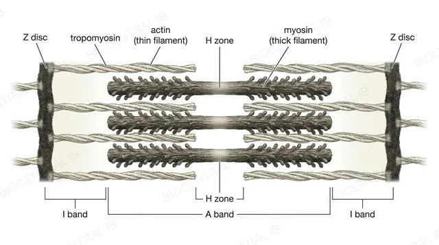
  • Within each sarcomere, thin actin filaments interleave with thick myosin filaments; the regulatory proteins troponin and tropomyosin sit on the actin filament, blocking the myosin-binding sites while the muscle is at rest.
Why it mattersEverything about how a muscle contracts happens at this one repeating unit — understand a single sarcomere and you understand the whole muscle.
A relaxed myofibril's sarcomere labeled exactly as in the IB data booklet: Z disc, actin (thin filament), myosin (thick filament), tropomyosin, I band, H zone, A band, clean scientific diagram
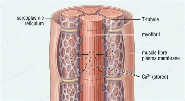
A muscle fibre cross-section with the sarcoplasmic reticulum wrapped around a myofibril storing calcium ions and a T-tubule carrying an electrical impulse inward, labeled
B3.3.2 · HL

Getting Ca²⁺ to exactly the right place

The sarcoplasmic reticulum and T-tubules form the delivery system that gets an electrical impulse and a burst of Ca²⁺ deep inside the muscle fibre almost instantly.
  • The sarcoplasmic reticulum, a specialised smooth ER wrapped around each myofibril, stores Ca²⁺ at high concentration, ready for release the instant a signal arrives.
  • T-tubules are inward folds of the muscle fibre's own plasma membrane, running close beside the sarcoplasmic reticulum; they carry the electrical impulse from the cell surface deep into the fibre's interior, reaching every sarcomere at once rather than only the ones near the edge.
Why it mattersWithout T-tubules, only sarcomeres near the cell surface would get the signal in time — the whole fibre needs to contract together.
B3.3.2 · HL

The sliding filament model, step by step

A motor neuron impulse triggers a precise chain of events that ends with actin and myosin filaments sliding past each other — the sliding filament model of contraction.
  • Acetylcholine release at the neuromuscular junction depolarises the fibre; the impulse travels down the T-tubules, and the sarcoplasmic reticulum releases Ca²⁺, which binds troponin and shifts tropomyosin aside to expose the myosin-binding sites on actin.
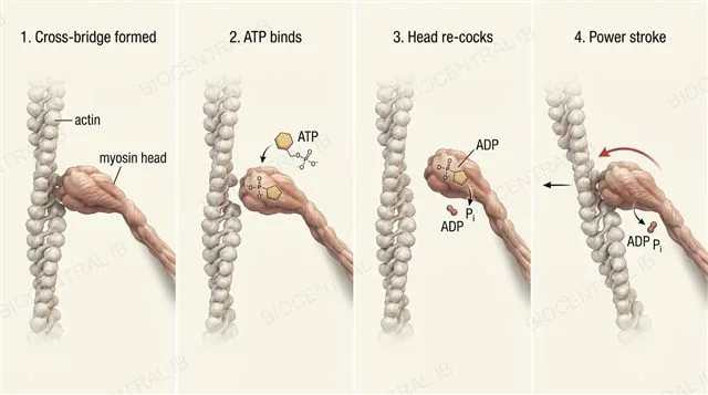
  • Myosin heads bind actin, forming cross-bridges; ATP binding breaks each cross-bridge and re-cocks the head, and the head's swivel — the power stroke — pulls actin past myosin. Repeated cycling ratchets the Z discs closer together, shortening the sarcomere without shortening either filament itself.
Why it mattersATP is needed to release the cross-bridge, not to form it — a common point of confusion worth remembering exactly.
The cross-bridge cycle: a myosin head binding actin, ATP breaking the cross-bridge and re-cocking the head, then the power stroke pulling actin past myosin, labeled sequence of four steps
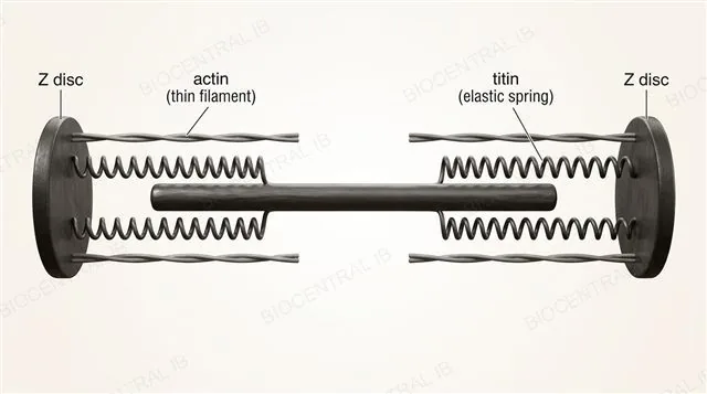
A stretched sarcomere with the elastic protein titin shown as a coiled spring running from the Z disc to the myosin filament, labeled
B3.3.3 · HL

Titin resets the sarcomere for free

Titin is an immense elastic protein running from the Z disc to the myosin filament, acting as a molecular spring within every sarcomere.
  • When a sarcomere is stretched, titin stores elastic potential energy, then uses it to help pull the sarcomere passively back to its resting length — no ATP required.
  • Titin also prevents overstretching, keeping the thick and thin filaments correctly aligned so cross-bridges can still form properly on the sarcomere's next contraction.
Why it mattersTitin's recoil is passive — it's the one part of muscle relaxation that costs the cell no energy at all.
B3.3.3 · HL

One muscle can only pull

Muscle tissue can only actively contract — it has no mechanism to actively push itself back out to a longer length.
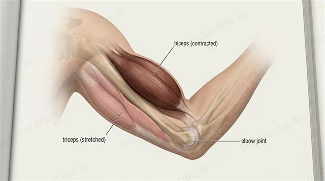
  • A muscle relaxing is never enough by itself to reverse its own shortening; antagonistic muscle pairs, such as the biceps and triceps at the elbow, solve this by having one member's contraction stretch the other back out.
  • When the biceps contracts to flex the arm, the triceps is stretched; when the triceps contracts to extend the arm, the biceps is stretched in turn — each contraction resets the other muscle's sarcomeres, ready for its own next contraction.
Why it matters"Antagonistic" doesn't mean fighting each other — it means taking turns doing the one thing muscle can do: pull.
The biceps and triceps as an antagonistic pair at the elbow, biceps contracted and shortened while triceps is stretched during arm flexion, labeled
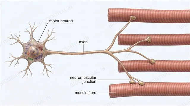
A single motor neuron branching to innervate several muscle fibres via neuromuscular junctions, labeled
B3.3.4 · HL

One neuron, every fibre it commands

A motor unit is a single motor neuron together with every muscle fibre it innervates — the smallest unit of controllable contraction.
  • The neuromuscular junction is the synapse between a motor neuron's terminal and a muscle fibre, where acetylcholine release triggers that fibre's own contraction sequence.
  • All fibres within one motor unit are innervated by branches of the same neuron, so they contract together as a single functional group whenever that neuron fires.
Why it mattersA muscle's contraction isn't one event — it's however many of its motor units happen to be firing at once.
B3.3.4 · HL

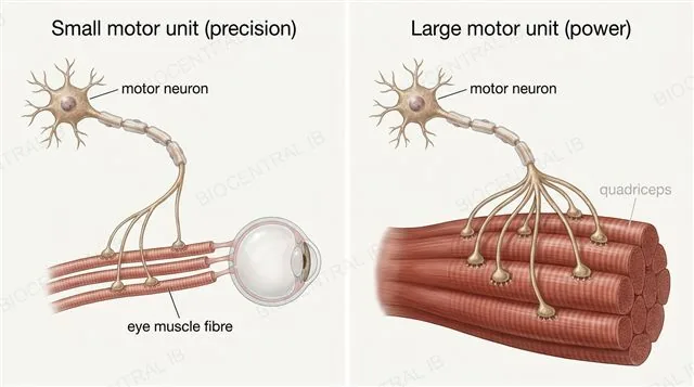
Motor unit size trades precision for power

The number of muscle fibres per motor neuron determines whether a muscle is built for fine control or for raw force.
  • Small motor units — few fibres per neuron, as in the muscles that move the eye — allow very fine, precise adjustments, since activating a single unit changes very little.
  • Large motor units — many fibres per neuron, as in the quadriceps — allow powerful contraction but coarser control, since activating a single unit moves a large mass of fibres at once.
Why it mattersThe eye needs precision far more than force; the quadriceps needs the opposite — motor unit size is matched to the job.
A small motor unit with a few muscle fibres controlling an eye compared with a large motor unit with many muscle fibres controlling the quadriceps, labeled precise versus powerful
Quick check · HL

A myosin head has just formed a cross-bridge with actin. ATP now binds to the myosin head. What does that ATP actually do at this point in the cycle?

It allows the myosin head to bind actin for the first time
It breaks the cross-bridge and re-cocks the myosin head, ready for the next power stroke
It triggers Ca²⁺ release from the sarcoplasmic reticulum
It powers troponin's shift away from the myosin-binding site
Correct answer: it breaks the cross-bridge and re-cocks the head. ATP binding releases myosin from actin and resets the head to its high-energy position. A common point of confusion is thinking ATP is needed for the initial binding step — that step doesn't require ATP at all.
Part two · HL

Skeletons, levers and joints

B3.3.5 – B3.3.8
B3.3.5 · HL

Arthropods anchor muscle to a rigid shell

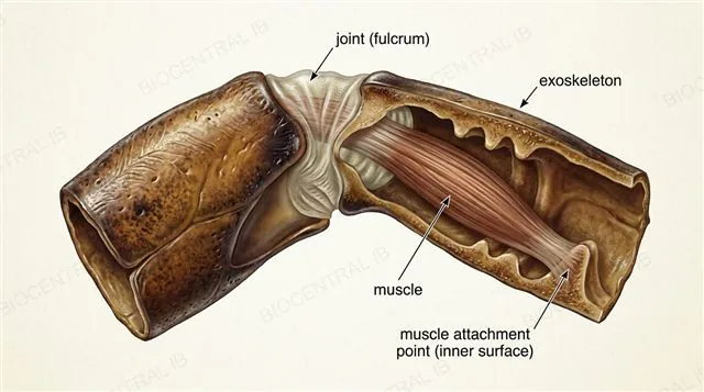
An exoskeleton is a rigid external casing, characteristic of arthropods, with muscles attached to its internal surface.
  • The exoskeleton provides fixed anchorage points on its inner surface for muscle attachment, letting muscles pull against a rigid frame rather than against soft tissue.
  • Joints between exoskeleton segments act as fulcrums, converting a muscle's pulling force into rotation of the limb segment beyond the joint — the skeleton functions as a lever system, not just armour.
Why it mattersAn exoskeleton does two jobs at once: protection on the outside, and a lever system for movement on the inside.
A cross-section of an arthropod leg joint showing the rigid exoskeleton, muscles attached to its internal surface, and the joint acting as a fulcrum, labeled, photorealistic detail
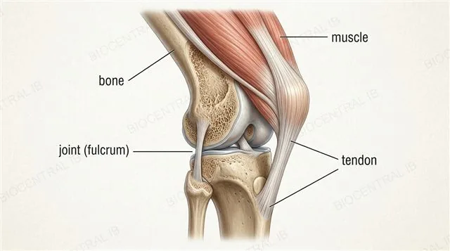
A cross-section of a vertebrate limb joint showing the internal endoskeleton, tendons attaching muscle to the outer bone surface, and the joint acting as a fulcrum, labeled
B3.3.5 · HL

Vertebrates anchor muscle from within

An endoskeleton is an internal skeleton, characteristic of vertebrates, with muscles attached externally via tendons.
  • Bones provide anchorage points on their outer surface for tendons to attach, and joints between bones act as fulcrums by the same lever principle as an exoskeleton.
  • Because the skeleton sits inside the body rather than around it, an endoskeleton can keep growing with the animal throughout life, unlike a rigid exoskeleton that must be periodically shed and regrown.
Why it mattersExo- and endoskeletons solve the same two problems — anchorage and leverage — from opposite sides of the body wall.
B3.3.6 · HL

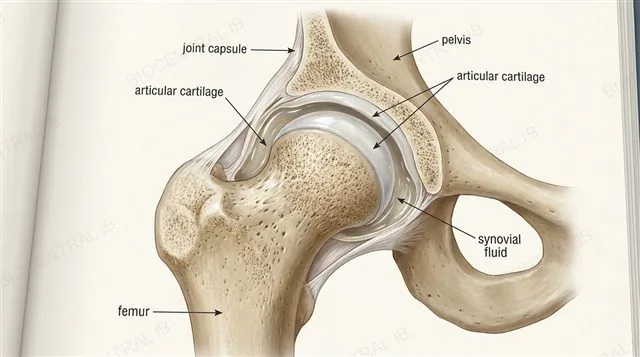
The hip: a joint built to move without wearing out

In a synovial joint such as the human hip, the femur and pelvis act as rigid levers either side of a capsule engineered to minimise friction.
  • Articular cartilage covers the bone surfaces where they meet, cushioning impact and reducing friction directly between the femur and pelvis.
  • Synovial fluid, held within the joint capsule, lubricates the joint further and helps absorb shock during movement and impact.
Why it mattersCartilage and synovial fluid together are why a joint used millions of times over a lifetime doesn't simply grind itself away.
A cross-section of the human hip synovial joint showing the femur, pelvis, articular cartilage and synovial fluid within the joint capsule, labeled
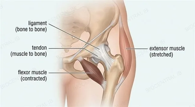
The human hip joint showing ligaments connecting femur to pelvis and tendons connecting muscle to bone, with flexor and extensor muscles crossing the joint as an antagonistic pair, labeled
B3.3.6 · HL

Ligaments hold it together, tendons make it move

Ligaments and tendons serve two distinct jobs at the synovial joint, and antagonistic muscles crossing it drive movement in opposite directions.
  • Ligaments connect bone to bone, stabilising the joint and limiting how far it can move; tendons connect muscle to bone, transmitting a contracting muscle's pulling force onto the lever.
  • Flexor and extensor muscles crossing the hip act as an antagonistic pair, exactly as at the elbow — one contracts to move the joint one way while being stretched back into position by the other.
Why it mattersA joint held together by ligaments alone wouldn't move at all — tendons are what turn a stable joint into a mobile one.
B3.3.7 · HL

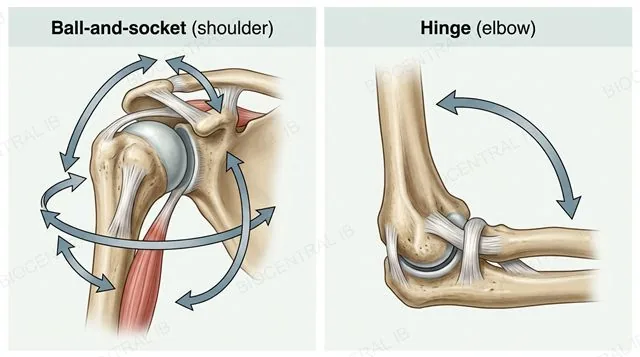
Not every joint moves the same way

Range of motion (ROM) is the angular distance and directions through which a joint can move, and it depends directly on the joint's shape.
  • Ball-and-socket joints, such as the hip and shoulder, allow movement in multiple planes at once — flexion, extension and rotation — giving them a large ROM.
  • Hinge joints, such as the elbow and knee, allow movement in essentially one plane only, flexion and extension, giving a smaller but more stable ROM. ROM can be measured directly with a goniometer or by computer analysis of motion.
Why it mattersA shoulder trades stability for range; a knee trades range for the stability it needs to bear weight — joint shape reflects that trade-off.
A ball-and-socket shoulder joint shown moving through multiple planes compared with a hinge elbow joint moving through one plane, with angle arcs labeled range of motion
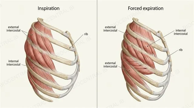
A ribcage cross-section showing external intercostal muscles contracted to raise the ribs and internal intercostal muscles stretched, labeled inspiration and expiration
B3.3.8 · HL

The ribcage moves by the same logic as a limb

The internal and external intercostal muscles, running between the ribs, apply the antagonistic-muscle principle to a movement that isn't a limb at all: breathing.
  • External intercostal muscles contract to raise the ribcage upward and outward, driving inspiration; internal intercostal muscles contract to pull it downward and inward, driving forced expiration.
  • Because the two muscle layers run in different fibre orientations, contracting one layer stretches the other, storing elastic potential energy in titin within the stretched muscle's own sarcomeres, ready for its turn to contract.
Why it mattersEven a movement with no obvious "joint" still runs on the same two ideas as a limb: antagonistic pairs, and titin's passive recoil.
Quick check · HL

A shoulder (ball-and-socket) has a far greater range of motion than a knee (hinge), but the knee is much more stable under load. Why?

The shoulder has more muscles attached to it than the knee
Ball-and-socket joints allow movement in multiple planes at once, while hinge joints move in essentially one plane — more planes of movement trades away stability
The knee has no synovial fluid, so it cannot move as freely
The shoulder is an exoskeleton joint and the knee is an endoskeleton joint
Correct answer: more planes of movement trades away stability. A ball-and-socket joint's freedom to rotate in multiple planes is exactly what makes it structurally less constrained — and therefore less stable — than a hinge joint restricted to one plane, which is why a weight-bearing joint like the knee favours the hinge design.
Part three · HL

Why animals move at all

B3.3.9 – B3.3.10
B3.3.9 · HL

Movement always serves a purpose

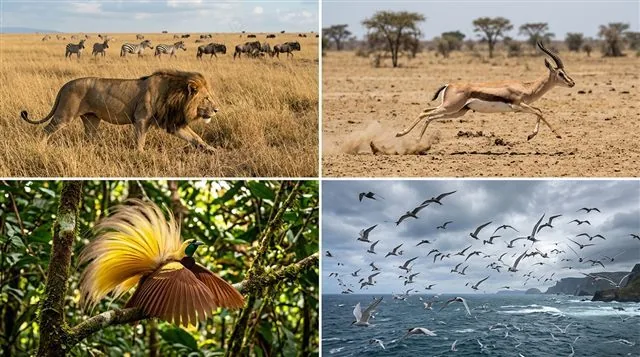
Locomotion is costly in energy, so it only evolves and persists where it serves one of a small number of clear purposes.
  • Foraging drives movement toward food, as when a lion stalks and pursues prey; escaping danger drives movement away from it, as when a gazelle flees a predator.
  • Finding a mate drives movement toward potential partners, as when male birds travel to display sites to court females; migration drives longer seasonal movement toward more favourable conditions, as when Arctic terns travel between hemispheres each year.
Why it mattersEvery case of locomotion in the exam is really answering the same question: what was this animal moving toward or away from, and why?
Four small scenes in one frame: a lion stalking prey, a gazelle fleeing, a bird displaying at a courtship site, and Arctic terns migrating, photorealistic wildlife photography style
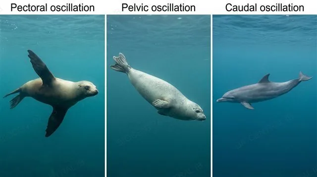
Three marine mammals compared: a sea lion sweeping front flippers, a true seal using hind flippers, and a dolphin beating its tail fluke up and down, labeled with propulsion mode names, photorealistic
B3.3.10 · HL

A body built to move through water, not air

Marine mammals show a consistent set of structural adaptations for swimming, starting with a streamlined body shape that reduces drag.
  • Limbs are modified into flippers or a tail fluke for propulsion and manoeuvring rather than walking.
  • Different groups use different propulsion modes: sea lions and fur seals use pectoral oscillation, sweeping their front flippers; true seals use hind-flipper (pelvic) oscillation; cetaceans such as whales and dolphins use caudal (tail) oscillation, moving the fluke up and down.
Why it mattersThree unrelated groups converged on flippers, but each still swims with a different stroke — the shared pressure was water, not a shared ancestor's technique.
B3.3.10 · HL

Breathing has to fit around swimming

Marine mammals also carry adaptations that let them breathe efficiently at the surface between dives.
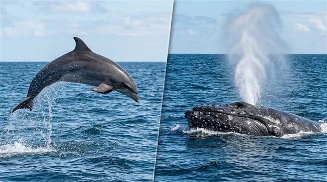
  • Nostrils positioned on top of the head, or modified into a blowhole, let an animal breathe with only a small part of its body breaking the surface.
  • Porpoising — leaping clear of the water while swimming — lets an animal breathe at speed with less drag than surfacing without leaving the water, useful when travelling or fleeing at full speed.
Why it mattersEvery adaptation here solves the same underlying problem: a lung-breathing animal has to keep swimming and keep breathing at the same time.
A dolphin porpoising, leaping clear of the water surface, and a whale's blowhole on top of its head releasing a breath spout, photorealistic

Key vocabulary

This entire topic is HL only — worth being able to define each in a single sentence

Sarcomere
The repeating contractile unit of a myofibril, bounded by Z discs.
Sliding filament model
Actin and myosin filaments sliding past each other, without shortening themselves, to contract a sarcomere.
Cross-bridge cycle
The repeating sequence of myosin binding actin, ATP releasing it, and the power stroke pulling the filaments.
Titin
An elastic protein linking myosin to the Z disc, providing passive elastic recoil.
Antagonistic muscles
A pair of muscles that act in opposition, one stretching the other as it contracts.
Motor unit
A motor neuron and every muscle fibre it innervates.
Range of motion (ROM)
The angular distance and directions through which a joint can move.
Synovial fluid
Lubricating fluid within a joint capsule that reduces friction and absorbs shock.
Porpoising
Leaping clear of the water while swimming, reducing drag at speed.

Where this shows up again

B2.2
The sarcoplasmic reticulum is a specialised form of smooth ER (B2.2). How does its Ca²⁺ storage and release function relate to the general role of SER in calcium homeostasis?
C1.2
ATP for muscle contraction is produced by aerobic respiration in mitochondria (C1.2). Explain why Type I fibres have more mitochondria than Type IIb, relating this to their different metabolic strategies.
C2.1
Ca²⁺ acts as a second messenger in both muscle contraction and cell signalling (C2.1). Compare the role of Ca²⁺ in triggering contraction with its role in neurotransmitter release at synapses.
C1.2
During intense exercise, muscles switch to anaerobic glycolysis producing lactate. How does this relate to the different ATP capacities of the three fibre types, and why does lactate accumulation limit performance?

B3.3 Muscle and motility — one-page recap

Screenshot this slide to revise from — this entire topic is HL only

Sarcomere & sliding filament
  • Muscle → fascicle → fibre → myofibril → sarcomere.
  • Actin + myosin slide; filaments don't shorten.
Excitation-contraction
  • SR stores Ca²⁺; T-tubules carry the impulse in.
  • ATP breaks the cross-bridge, not forms it.
Titin & antagonistic pairs
  • Titin recoils the sarcomere passively.
  • Muscle only pulls — needs a partner to reset.
Motor units
  • Small = precision (eye); large = power (quads).
  • Neuron + all fibres it innervates.
Skeletons & joints
  • Exo- vs endoskeleton: anchorage + lever.
  • Synovial joint; ball-socket vs hinge ROM.
Why & how animals move
  • Forage, flee, mate, migrate.
  • Marine mammals: streamlining, flippers, blowhole.

Every movement starts with one pull

A sarcomere can only shorten — skeletons, joints and antagonistic pairs are what turn that single motion into a sprint, a dive, or a breath.
B3.3 Muscle and motility · BioCentral IB
Use ↓ ↑ or click to navigate
01 / 22