BioCentral IBBioCentral IB
IB Biology · Theme C · C1.1

Enzymes and metabolism

From a single active site cradling its substrate to the thousands of linked reactions that keep a cell alive, this lesson traces how enzymes make metabolism fast enough, specific enough, and controllable enough to sustain life.
Guiding questions

In what ways do enzymes interact with other molecules?

What are the interdependent components of metabolism?

Part one

Enzymes as catalysts

C1.1.1 – C1.1.7
C1.1.1

Speeding up life's essential chemistry

Enzymes are biological catalysts: proteins that speed up chemical reactions in cells without being consumed or permanently changed in the process.
  • Left to random collisions alone, most of the reactions life depends on would be far too rare to sustain a cell — enzymes lower the energy barrier so useful chemistry happens on a timescale of milliseconds rather than years.
  • Because an enzyme is released unchanged at the end of each reaction, one enzyme molecule can catalyse the same reaction thousands of times over, so only a small quantity is needed to keep a pathway running continuously.
Why it mattersRapid energy release, fast waste removal, and quick homeostatic responses such as blood clotting are only possible because enzymes make these reactions fast enough to matter.
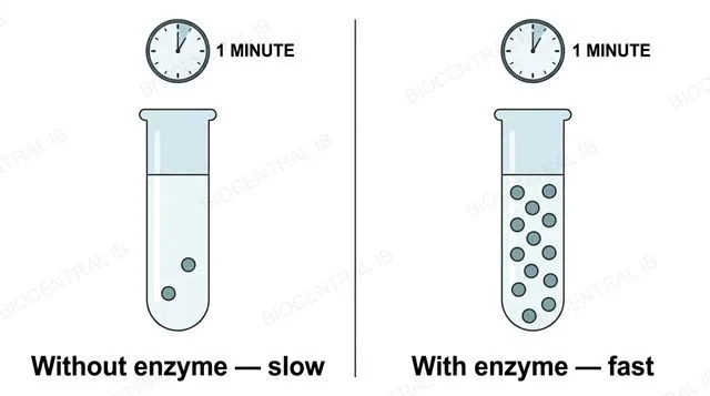
Side-by-side comparison diagram showing very few product molecules forming without an enzyme present versus many product molecules forming quickly with an enzyme present, labeled without enzyme and with enzyme
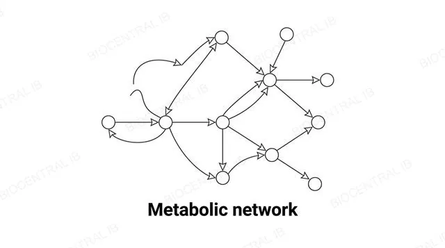
A branching network diagram of interconnected metabolic pathways, nodes representing metabolites connected by arrows representing enzyme-catalysed reactions, labeled metabolic network
C1.1.2

One cell, thousands of enzymes

Metabolism is the complex network of interdependent, interacting chemical reactions occurring inside a living organism, and enzymes are what make that network function.
  • Because each enzyme's active site is specific to a particular substrate (or a small family of related substrates), a cell needs a distinct enzyme for nearly every different reaction — which is why genomes encode thousands of different enzymes.
  • The product of one enzyme-catalysed reaction typically becomes the substrate for the next enzyme in a pathway, linking individual reactions into long interdependent chains and cycles rather than isolated events.
Why it mattersControlling which enzymes are active — switching them on, off, up or down — is how a cell exerts control over its entire metabolism without rewriting its DNA.
C1.1.3

Building up, breaking down

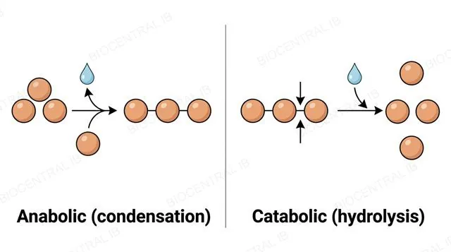
Anabolic reactions build larger molecules from smaller ones and generally require energy input, while catabolic reactions break larger molecules down and generally release energy.
  • Anabolism proceeds by condensation reactions that join monomers into macromolecules — protein synthesis from amino acids, glycogen formation from glucose, and the sugar-building reactions of photosynthesis are all examples.
  • Catabolism proceeds by hydrolysis, using water to break bonds — digestion splitting macromolecules into monomers, and the oxidation of substrates during cell respiration, are the guide's named examples.
Why it mattersEvery metabolic pathway in this topic — respiration, photosynthesis, digestion — is really just anabolism or catabolism happening through a specific sequence of enzymes.
Comparison diagram: small monomer molecules joining together into a larger polymer chain with a water molecule released, labeled anabolic condensation, next to a larger polymer chain breaking apart into monomers with a water molecule added, labeled catabolic hydrolysis
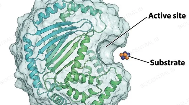
A detailed enzyme molecule rendered as a folded globular protein with a visible pocket-shaped active site on its surface, a small substrate molecule approaching the pocket, labeled active site and substrate
C1.1.4

A pocket built from folding

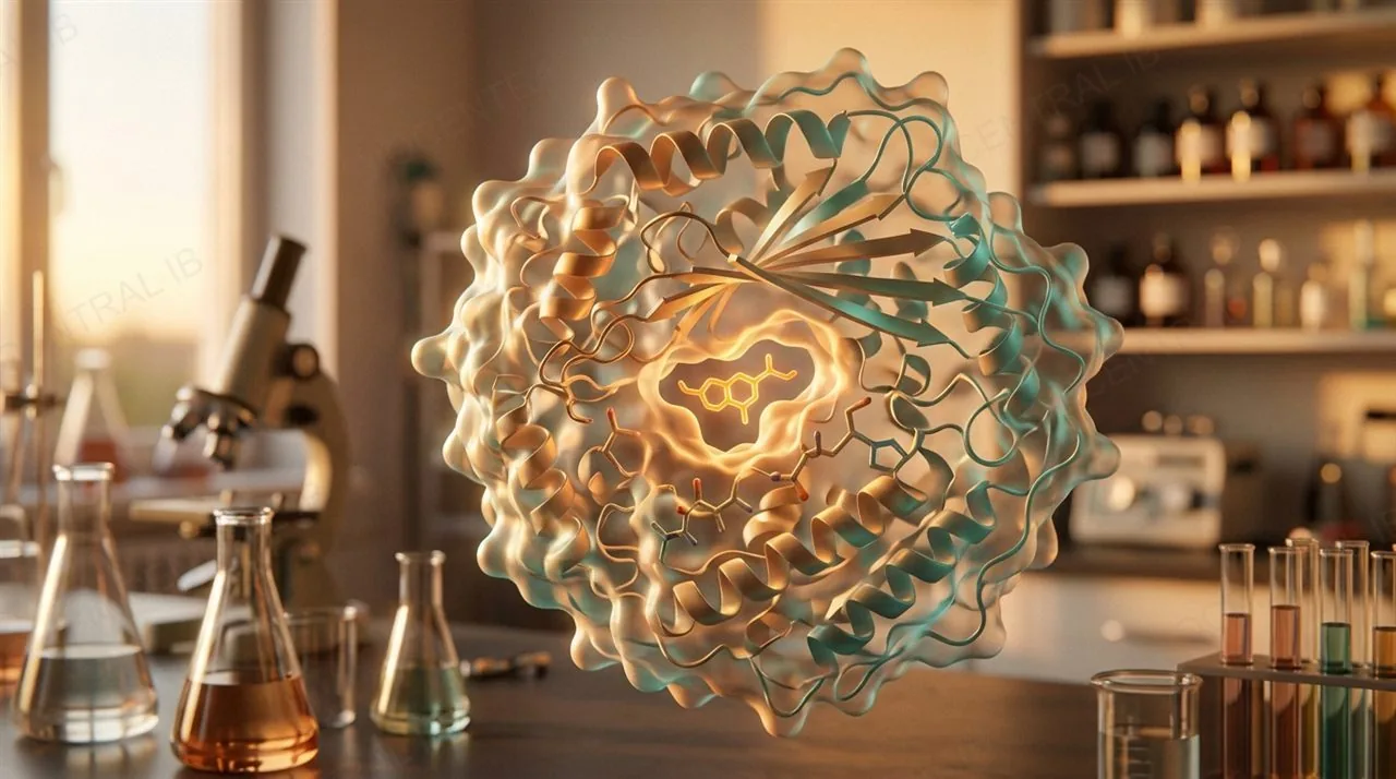
Enzymes are globular proteins whose three-dimensional folding creates a small pocket or cleft, the active site, where catalysis actually happens.
  • The active site itself is formed by only a few amino acids, but it is the folding of the enzyme's entire tertiary structure that positions those few amino acids correctly relative to each other.
  • Interactions between amino acids across the whole protein — not just those lining the pocket — hold the active site in the precise shape and chemical environment that catalysis requires.
Why it mattersThis is why even a mutation far from the active site can still destroy an enzyme's function — it can distort the folding that positions the active site correctly.
C1.1.5

Substrate and enzyme both change shape

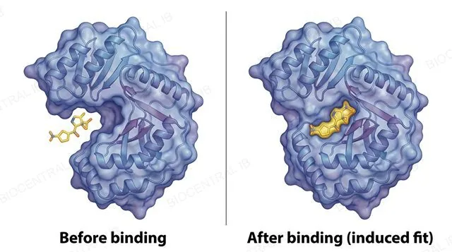
In the induced-fit model, the active site is already a broadly complementary match for its substrate, and binding triggers a small conformational change in both molecules that tightens the fit.
  • Before binding, the active site's shape is an approximate match; binding refines that fit and correctly aligns the catalytic groups needed for the reaction, rather than the enzyme having no shape at all beforehand.
  • The substrate itself also changes shape slightly during binding — this strains particular bonds within it, helping to push the substrate toward its high-energy transition state.
Why it mattersInduced fit replaced the older, more rigid lock-and-key model because it explains why enzymes can still act on substrates that are only an approximate — not identical — shape match.
Two-panel diagram of the same enzyme active site, left panel labeled before binding showing an approximately shaped pocket near a substrate, right panel labeled after binding (induced fit) showing the pocket slightly reshaped tightly around the substrate
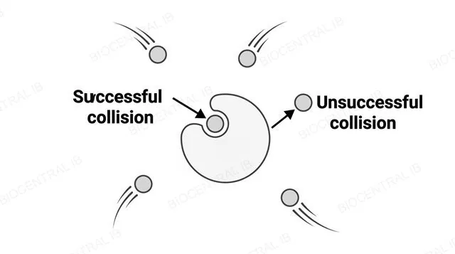
A diagram of enzyme and substrate molecules scattered in solution with curved motion-trail arrows showing random undirected movement, one substrate colliding successfully into an active site labeled successful collision, another substrate bouncing off at the wrong angle labeled unsuccessful collision
C1.1.6

Catalysis starts with a collision

Enzymes and substrates dissolved in a cell meet through random thermal (Brownian) motion, not through any directed movement toward each other.
  • Both molecules are in constant, undirected motion due to their kinetic energy, so an enzyme-substrate collision happens purely by chance — only a fraction of collisions occur with the correct orientation and enough energy to form a complex.
  • Because movement is required to bring substrate and active site together, anything that increases collision frequency — higher temperature, higher concentration — tends to increase the reaction rate, up to a point.
Why it mattersThis randomness is exactly why enzyme kinetics can be described statistically, as in the upcoming rate graphs, even though any single collision is unpredictable.
C1.1.6

When something can't move

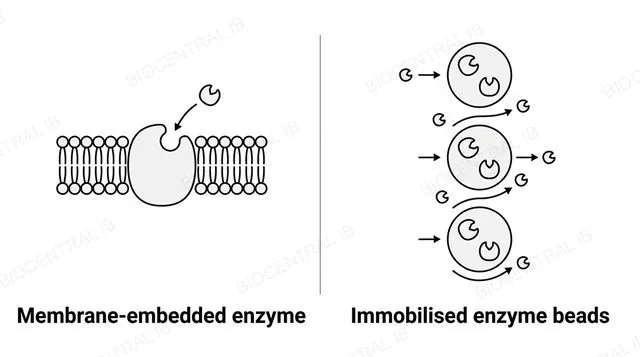
Molecular motion isn't always free: some large substrate molecules are effectively immobilised, and some enzymes are immobilised too, either naturally or industrially.
  • Naturally, some enzymes are immobilised by being embedded directly in a membrane, so the enzyme stays fixed in place while smaller, mobile substrate molecules diffuse to reach it.
  • Industrially, enzymes such as lactase are deliberately immobilised — trapped in alginate beads or bound to an inert surface — so they can be recovered, reused, and kept separate from the product, as in lactose-free milk production.
Why it mattersImmobilisation doesn't make an enzyme catalyse faster; it makes the process more practical and cost-effective to run at scale.
Two labeled panels: left panel showing an enzyme molecule embedded fixed within a cell membrane with a small substrate molecule diffusing toward it, labeled membrane-embedded enzyme; right panel showing spherical alginate beads containing trapped enzyme molecules with substrate flowing past in a column, labeled immobilised enzyme beads
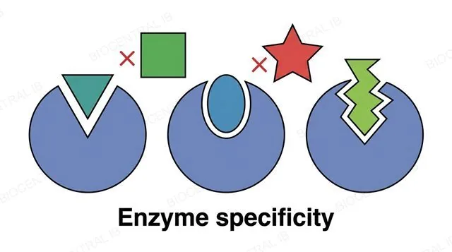
Three differently shaped enzyme active site pockets each shown with one complementary substrate fitting snugly into its matching pocket, and two non-matching substrates shown unable to fit into the wrong pockets, labeled enzyme specificity
C1.1.7

Why one enzyme won't touch another substrate

Enzyme-substrate specificity — an enzyme only catalysing a reaction with a particular substrate or family of related substrates — comes directly from the exact structure of the active site.
  • The active site's shape and chemical environment, its mix of hydrophobic, charged and polar R groups, are set by the enzyme's tertiary structure, so only a substrate complementary in both shape and chemistry can bind effectively.
  • Two enzymes sitting in the very same organelle can have completely different specificities, because specificity depends on active-site structure, not on where an enzyme happens to be located.
Why it mattersSpecificity is what lets a cell run thousands of separate reactions side by side without them interfering with each other.
C1.1.7

Denaturation: same sequence, wrong shape

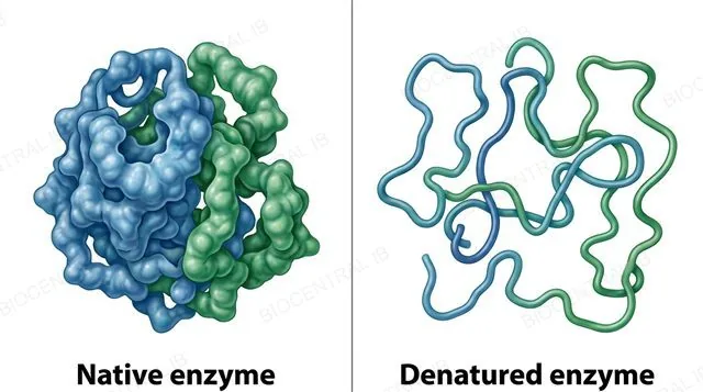
Denaturation is a usually irreversible change to an enzyme's tertiary structure that distorts or destroys its active site, without breaking the primary sequence of amino acids itself.
  • Extreme temperature or pH disrupts the hydrogen bonds and ionic interactions holding the tertiary structure together, so the protein unfolds and the active site loses the precise shape substrate binding depends on.
  • The covalent peptide bonds of the primary structure are not broken — the amino acid sequence is unchanged — which is why denaturation is a shape problem, not a chemical-composition problem.
Why it mattersOnce an active site is denatured, the substrate can no longer bind properly, so catalysis stops even though the enzyme molecule is still physically present.
Two-panel diagram: left panel labeled native enzyme showing a tightly folded globular protein with an intact pocket-shaped active site; right panel labeled denatured enzyme showing the same protein chain unfolded and tangled with the active site pocket visibly distorted and gone
Quick check

Two enzymes are found in the exact same cell compartment but catalyse completely different reactions. What explains this difference in specificity?

They must actually be located in different organelles for this to be possible
Their active sites have different shapes and chemical environments, set by each enzyme's own tertiary structure
One of the two enzymes has already been denatured
Enzymes sharing a compartment always catalyse the same reaction
Correct answer: different active-site structure. Specificity depends entirely on the precise shape and chemistry of an enzyme's active site — a property of its tertiary structure, not its location. Two enzymes can share a compartment and still have entirely different specificities.
Part two

Enzyme kinetics

C1.1.8 – C1.1.10
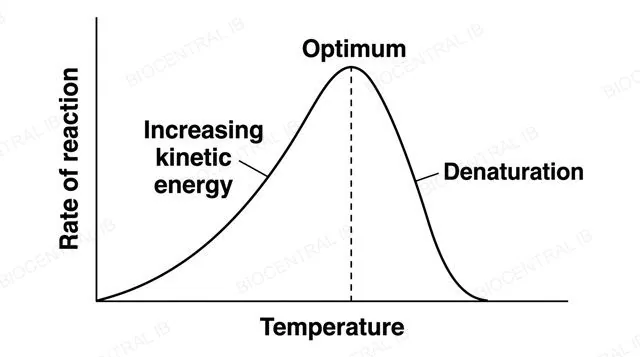
A line graph of reaction rate on the y axis against temperature on the x axis, showing rate rising smoothly, peaking at a labeled optimum temperature, then falling sharply, with the rising portion labeled increasing kinetic energy and the falling portion labeled denaturation
C1.1.8

Rate rises, then collapses

Reaction rate increases with temperature up to an optimum, then falls sharply as the enzyme denatures — two different effects producing one curve.
  • Below the optimum, rising temperature increases the kinetic energy of enzyme and substrate molecules, producing more frequent, more energetic collisions and therefore a faster rate.
  • Beyond the optimum, rising temperature increasingly breaks the weak bonds holding tertiary structure together, denaturing a growing fraction of the enzyme population, so rate falls sharply even as substrate remains plentiful.
Why it mattersA generalised sketch graph like this is a model in biology — real experimental data is what tests how well the sketch matches the actual relationship.
C1.1.8

Every enzyme has its own optimum pH

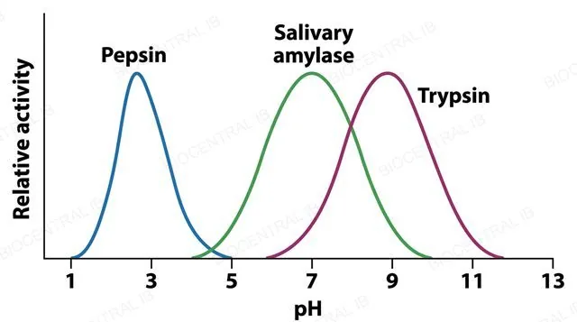
Enzyme activity peaks at a specific optimum pH and falls away on either side, because pH changes the ionisation of active-site R groups and can denature the protein.
  • Most human enzymes function best near neutral pH, matching the cytoplasm, but exceptions exist: pepsin's optimum is around pH 2 in the stomach, while trypsin's is around pH 8 in the small intestine.
  • Away from the optimum, altered R-group charges disrupt substrate binding, and extreme pH also breaks the ionic and hydrogen bonds maintaining tertiary structure, denaturing the enzyme.
Why it mattersAn enzyme's optimum pH is a biochemical adaptation to the specific environment it normally works in.
A line graph of relative enzyme activity on the y axis against pH from 1 to 13 on the x axis, showing three separate bell-shaped curves, one peaking sharply near pH 2 labeled pepsin, one peaking near pH 7 labeled salivary amylase, and one peaking near pH 8 labeled trypsin
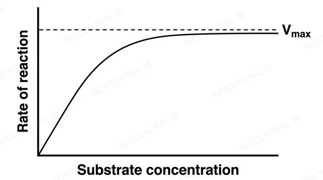
A line graph of reaction rate on the y axis against substrate concentration on the x axis, showing rate rising steeply at low concentration then levelling off into a flat plateau, with a dashed horizontal line marking the plateau labeled Vmax
C1.1.8

Why rate plateaus at Vmax

As substrate concentration rises, reaction rate increases until it levels off at Vmax, the maximum rate the enzyme population can sustain.
  • At low substrate concentration, not every active site is occupied at a given moment, so adding more substrate increases the rate almost linearly by increasing successful collisions.
  • At saturating substrate concentration, every active site is occupied continuously, so the rate is limited only by how fast each site can process substrate — adding still more substrate cannot push the rate any higher.
Why it mattersPast Vmax, only adding more enzyme — not more substrate — can increase the rate further.
C1.1.9

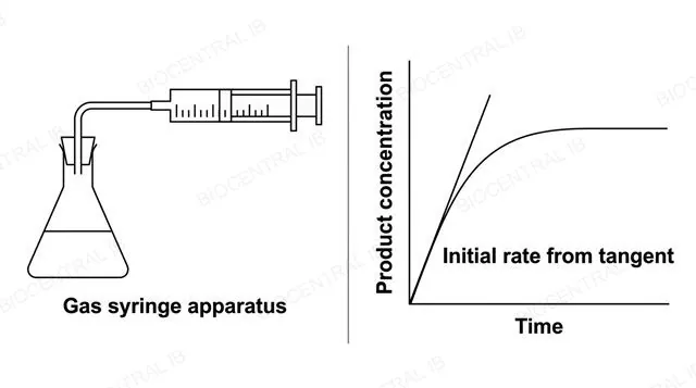
Turning a curve into a number

Reaction rate can be determined experimentally either continuously, by tracking product formation over time, or as a single fixed endpoint.
  • For continuous data — a graph of product concentration rising then levelling off — the initial rate is found by drawing a tangent to the curve at t = 0 and calculating its gradient, before substrate depletion slows the reaction.
  • When only a single endpoint can be timed, such as a solution turning cloudy, rate is calculated as 1 ÷ time taken, since a shorter time to the endpoint means a faster reaction.
Why it mattersApparatus should match what's actually being measured — a gas syringe for a gas product, a colorimeter for a colour change — using the wrong tool measures the wrong quantity.
Two-panel diagram: left panel showing a labeled gas syringe apparatus connected to a reaction flask collecting gas produced during an enzyme-catalysed reaction; right panel showing a graph of product concentration over time rising then levelling off with a straight tangent line drawn touching the curve at time zero, labeled initial rate from tangent
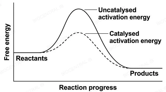
An energy profile graph with free energy on the y axis and reaction progress on the x axis, showing reactants and products at the same two energy levels for both curves, one curve with a tall hump labeled uncatalysed activation energy and a second lower curve with a shorter hump labeled catalysed activation energy
C1.1.10

Lowering the barrier, not the drop

Activation energy (Eₐ) is the minimum energy needed to destabilise bonds in the substrate enough to reach the high-energy transition state from which a reaction can proceed.
  • Enzymes lower Eₐ by binding substrate in the correct orientation, straining specific bonds, and stabilising the transition state itself — none of which requires adding external energy to the substrate.
  • Both the catalysed and uncatalysed pathway start and end at the same reactant and product energy levels, so the overall free-energy change (ΔG) of the reaction is completely unchanged by the enzyme.
Why it mattersOnly the height of the energy barrier changes, not how much energy the reaction ultimately releases or requires — a common exam-trap distinction.
Part three · HL

Metabolism as an interconnected network

C1.1.11 – C1.1.13
C1.1.11 · HL

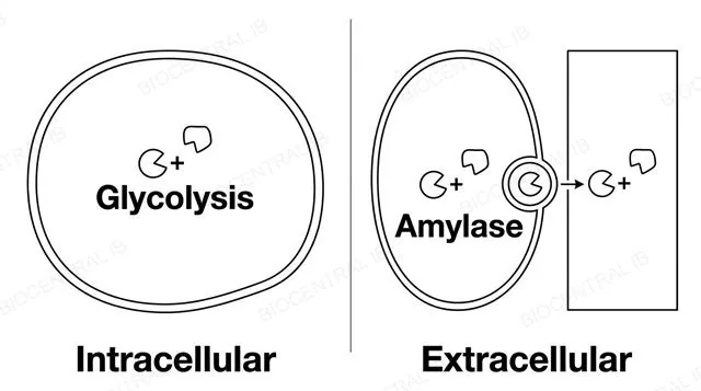
Inside the cell, or outside it

Intracellular enzymes catalyse reactions inside the same cell that made them, while extracellular enzymes are secreted to act outside that cell.
  • Glycolysis (cytoplasm) and the Krebs cycle (mitochondrial matrix) are intracellular examples — being inside a specific organelle doesn't make an enzyme extracellular, since "intracellular" just means acting inside the producing cell.
  • Chemical digestion in the gut is the guide's extracellular example: enzymes such as amylase are made in secretory cells, packaged into vesicles, and released by exocytosis into the gut lumen, which is topologically outside the body's own cells.
Why it mattersThis distinction connects directly to condensation and hydrolysis reactions (C1.1.3) and to digestion elsewhere in the course, which depends on these extracellular reactions having already happened.
Two-panel diagram: left panel showing a cell with an enzyme labeled glycolysis working inside its own cytoplasm on an internal substrate, labeled intracellular; right panel showing a secretory cell releasing an enzyme labeled amylase by exocytosis into a tube-shaped gut lumen outside the cell, labeled extracellular
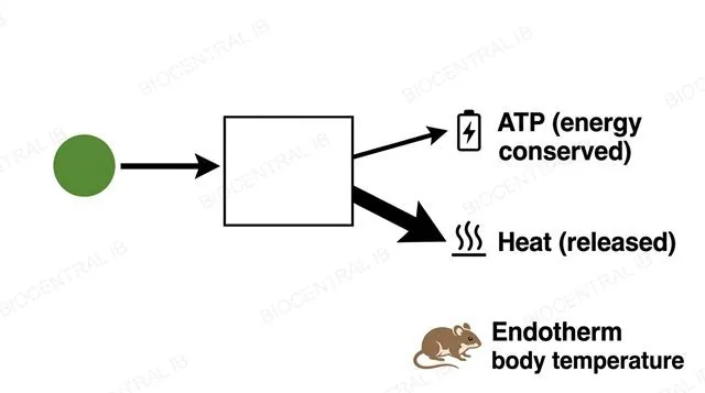
A flow diagram showing chemical energy from a substrate entering a metabolic reaction and splitting into two labeled output arrows, a smaller arrow labeled ATP energy conserved and a larger arrow labeled heat released, next to a small mammal icon labeled endotherm body temperature
C1.1.12 · HL

Metabolism is never perfectly efficient

Heat generation by metabolic reactions is inevitable, because no biological energy transfer is 100% efficient — some energy is always released as heat rather than conserved.
  • During reactions such as those of cell respiration, only part of the energy released is captured in ATP; the rest is unavoidably lost as heat, regardless of how well-adapted the enzyme is.
  • Mammals, birds and some other animals rely on this inevitable heat output, combined with a comparatively high metabolic rate, to maintain a stable, elevated body temperature.
Why it mattersShivering is this principle in action: contracting muscle raises the rate of respiration, and the resulting heat — not a direct heating mechanism — warms the body.
C1.1.13 · HL

Some pathways loop back to the start

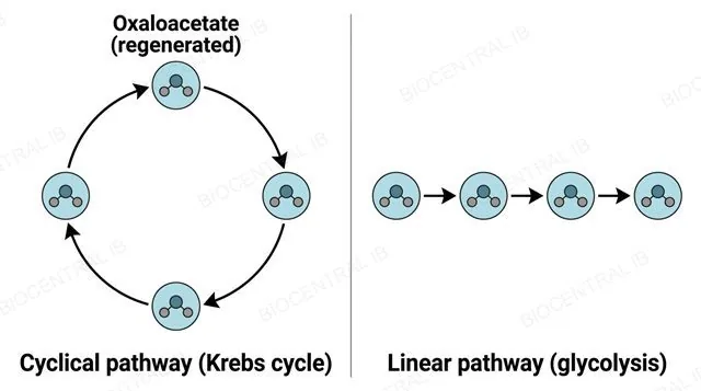
A cyclical pathway regenerates its starting molecule, or a key intermediate, at the end of each turn, while a linear pathway proceeds one-way to a final product without regenerating anything.
  • The Krebs cycle regenerates oxaloacetate and the Calvin cycle regenerates RuBP (ribulose bisphosphate) at the end of each turn, allowing the same molecules to combine with new substrate and begin again.
  • Glycolysis is the guide's linear example: glucose proceeds through a fixed sequence of steps to pyruvate without any starting molecule being regenerated to restart the sequence.
Why it mattersHaving several enzyme-catalysed steps doesn't make a pathway cyclical — only regeneration of the initial substrate or intermediate does.
Two-panel diagram: left panel showing four molecules connected in a closed circular arrow loop labeled cyclical pathway (Krebs cycle) with oxaloacetate regenerated at the starting point; right panel showing four molecules connected by a straight one-directional arrow sequence labeled linear pathway (glycolysis) ending at a final product with no loop back
Part four · HL

Regulating enzyme activity

C1.1.14 – C1.1.17
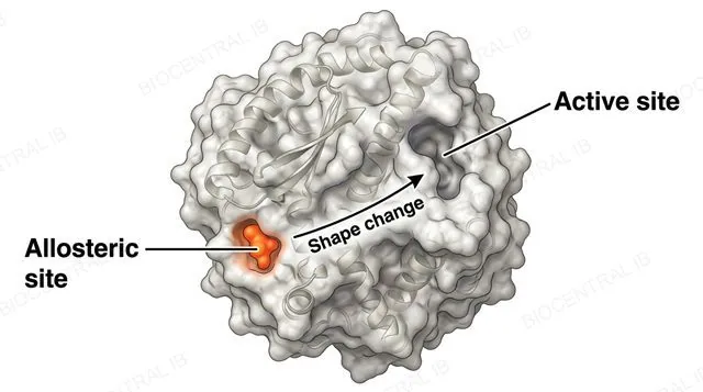
An enzyme molecule diagram showing two distinct labeled sites, a pocket-shaped active site on one part of the surface and a separate allosteric site elsewhere on the surface with an inhibitor molecule bound there, with an arrow showing the resulting shape change distorting the active site
C1.1.14 · HL

A second site, far from the active site

An allosteric site is a structurally distinct regulatory site, separate from the active site, where only specific substances can bind.
  • Binding at the allosteric site causes conformational changes that propagate through the enzyme's structure, altering the shape of the active site enough to prevent catalysis, even though the inhibitor never enters the active site itself.
  • This binding is reversible — the inhibitor can detach again, restoring the active site's original shape and the enzyme's normal activity.
Why it mattersNon-competitive inhibition works by remote control: distorting the active site indirectly, rather than blocking it directly the way a competitive inhibitor does.
C1.1.15 · HL

Competitive vs non-competitive

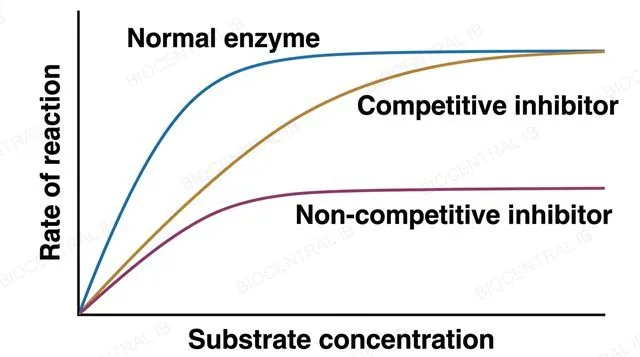
A competitive inhibitor binds reversibly to the active site itself, competing directly with the substrate; statins are the guide's example, competitively inhibiting HMG-CoA reductase to lower cholesterol synthesis.
Competitive
  • Binds the active site, resembling the real substrate
  • Overcome by raising [substrate] — enough substrate outcompetes the inhibitor
  • Vmax unchanged; Km increases
Non-competitive
  • Binds the separate allosteric site, not the active site
  • NOT overcome by raising [substrate] — the inhibitor isn't competing for that site
  • Vmax decreases; Km unchanged
Why it mattersStatins are a reversible, competitive inhibitor — the guide's own named example, not just a generic case study.
A line graph of reaction rate on the y axis against substrate concentration on the x axis, showing three curves that all start at the origin: an uninhibited enzyme curve reaching the highest plateau, a competitively inhibited curve rising more slowly but reaching the same plateau, and a non-competitively inhibited curve reaching a distinctly lower plateau, each curve labeled by name
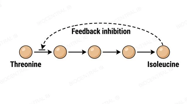
A linear metabolic pathway diagram showing threonine converted through several labeled intermediate steps into isoleucine at the end, with a curved dashed arrow looping back from the isoleucine end product to inhibit the first enzyme in the pathway, labeled feedback inhibition
C1.1.16 · HL

The end product shuts off its own pathway

In feedback inhibition, the end product of a metabolic pathway binds to an allosteric site on an enzyme early in that same pathway, reducing its activity.
  • The guide's named example is the pathway producing isoleucine from threonine: once enough isoleucine has accumulated, it binds allosterically to the first enzyme in the pathway and inhibits it.
  • Because this shuts down the pathway at its very first committed step, feedback inhibition prevents the cell from wastefully overproducing a product it no longer needs.
Why it mattersFeedback inhibition is reversible — as isoleucine is used up elsewhere, its concentration falls, the enzyme is released from inhibition, and production resumes automatically.
C1.1.17 · HL

An inhibitor that never lets go

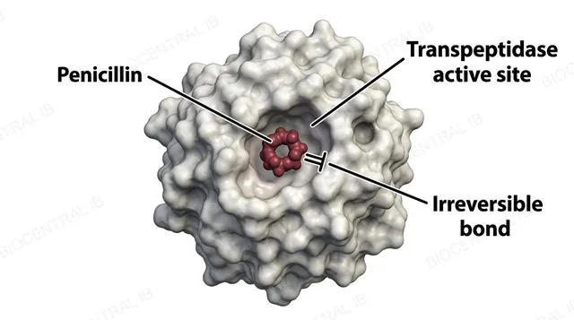
Mechanism-based inhibition occurs when an inhibitor binds to the active site and causes an irreversible chemical change, permanently inactivating the enzyme.
  • Penicillin is the guide's named example: it binds to bacterial transpeptidase enzymes, which normally cross-link peptidoglycan during cell wall synthesis, and causes an irreversible chemical change to their active site.
  • Because the enzyme can never return to its original state, one molecule of a mechanism-based inhibitor permanently removes one enzyme molecule from the active pool, unlike the reversible binding of competitive or non-competitive inhibitors.
Why it mattersThis irreversibility is exactly why some bacteria evolve resistance, by producing altered transpeptidases that penicillin can no longer bind or modify.
A diagram showing a penicillin molecule binding into the active site of a bacterial transpeptidase enzyme, with a labeled arrow showing an irreversible chemical bond forming permanently between the inhibitor and the active site, labeled mechanism-based inhibition
Quick check · HL

An inhibitor binds reversibly at a site distinct from an enzyme's active site, and raising substrate concentration does not restore normal activity. What type of inhibition is this?

Non-competitive inhibition — binding at the allosteric site distorts the active site without competing for it
Competitive inhibition — the inhibitor is simply outcompeting the substrate for the active site
Mechanism-based inhibition — the enzyme has been permanently and irreversibly inactivated
Feedback inhibition — the inhibitor must be the pathway's own end product
Correct answer: non-competitive inhibition. Reversible binding away from the active site, with no rescue from added substrate, is the defining signature of non-competitive (allosteric) inhibition — competitive inhibition is overcome by excess substrate, and mechanism-based inhibition is irreversible.

Key vocabulary

Worth being able to define in a single sentence each

Active site
The region of an enzyme, formed by its 3D folding, where a substrate binds and catalysis occurs.
Induced fit
Model in which substrate binding triggers a small conformational change that improves the fit between enzyme and substrate.
Denaturation
A usually irreversible change to a protein's tertiary structure that destroys its function without breaking its primary sequence.
Metabolite
Any molecule taking part in the reactions of metabolism, including substrates, intermediates and products.
Activation energy
The minimum energy needed to reach a reaction's transition state; enzymes lower it without changing ΔG.
Vmax
The maximum rate an enzyme population can sustain, reached once every active site is continuously occupied.
Allosteric site HL
A regulatory binding site distinct from the active site, where only specific effector molecules bind.
Feedback inhibition HL
Regulation in which a pathway's end product inhibits an enzyme early in that same pathway.
Mechanism-based inhibition HL
Irreversible inactivation caused by an inhibitor chemically modifying the active site.

Where this shows up again

B1.2
The tertiary structure of an enzyme (B1.2) creates its active site. Using a named enzyme as an example, explain how the specific R groups in the active site contribute to substrate binding and catalysis.
B2.2
Lysosomes (B2.2) contain hydrolytic enzymes that function optimally at pH ~5. Explain why these enzymes must be compartmentalised, and what would happen if they leaked into the cytoplasm (pH ~7.2).
C1.2
The energy for anabolic reactions is provided by ATP hydrolysis. Explain how the ATP produced during cell respiration (C1.2) is coupled to endergonic reactions via enzyme-catalysed phosphorylation.
C1.2
During germination, starch in seeds is hydrolysed by amylase into glucose, which is then respired. Explain why amylase activity increases during germination, linking this to the metabolic demands of the growing seedling.

C1.1 Enzymes and metabolism — one-page recap

Screenshot this slide to revise from

Enzymes as catalysts
  • Enzymes lower activation energy without being consumed; ΔG unchanged.
  • Active site shape, from tertiary structure, drives specificity.
Induced fit & denaturation
  • Substrate binding induces a small conformational change in both molecules.
  • Denaturation distorts the active site irreversibly; primary structure stays intact.
Enzyme kinetics
  • Rate rises then collapses with temperature (collision + denaturation).
  • Rate plateaus at Vmax once every active site is saturated.
HL · Metabolism as a network
  • Anabolism (condensation, energy in) vs catabolism (hydrolysis, energy out).
  • Intracellular (glycolysis, Krebs) vs extracellular (gut digestion).
HL · Cycles & heat
  • Krebs/Calvin cycles regenerate their starting molecule; glycolysis doesn't.
  • Heat is an inevitable, non-100%-efficient byproduct of metabolism.
HL · Inhibition
  • Competitive (active site, reversible, statins) vs non-competitive (allosteric, reversible).
  • Feedback (isoleucine) reversible; mechanism-based (penicillin) irreversible.

Life's chemistry, catalysed and controlled

From one folded active site to an entire interdependent network, every reaction in this lesson comes down to the same idea: shape determines function.
C1.1 Enzymes and metabolism · BioCentral IB
Use ↓ ↑ or click to navigate
01 / 33