BioCentral IBBioCentral IB
IB Biology · Theme C · C2.1 · HL only

Chemical signalling

From a hormone crossing the width of the body to a G protein switching on in a fraction of a second, this lesson traces how cells build, send and interpret the chemical messages that coordinate an entire animal body.
Guiding questions

How do cells distinguish between the many different signals that they receive?

What interactions occur inside animal cells in response to chemical signals?

Part one

Signalling chemicals and receptors

C2.1.1 – C2.1.5
C2.1.1

A protein shaped to fit one signal

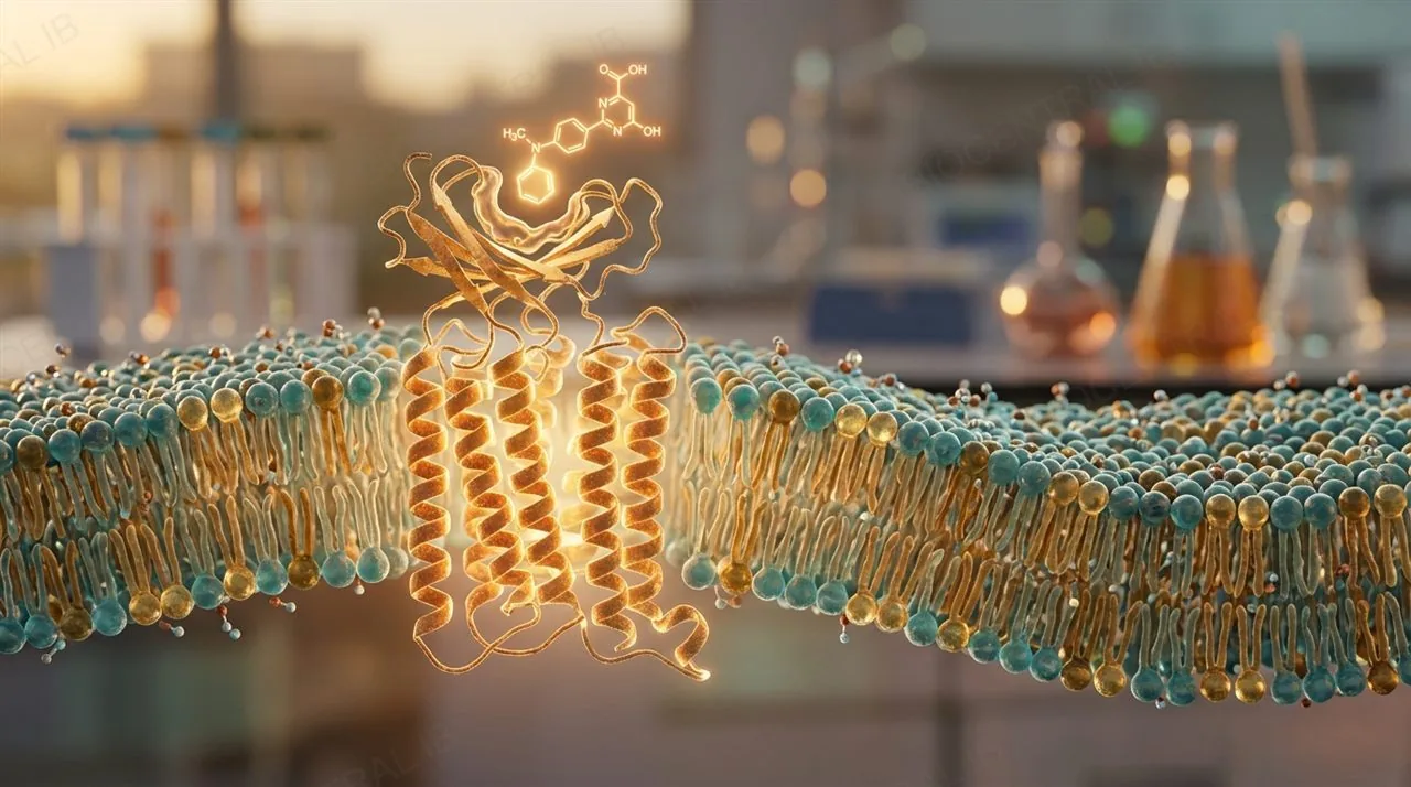
A receptor is a protein with a binding site whose shape and chemistry are complementary to a specific signalling chemical — the IB syllabus term for this signalling chemical is a ligand.
  • A receptor's binding site only accepts a ligand, or a small family of closely related ligands, whose shape and chemical groups match it — in the same way an enzyme's active site is complementary to its substrate.
  • Binding triggers a conformational change in the receptor, a shift in its three-dimensional shape, and it is this shape change — not the destruction of either molecule — that starts the cell's response.
Why it mattersA receptor is reused, not consumed: once its ligand detaches or is broken down, the receptor returns to its original shape and becomes available to bind another ligand.
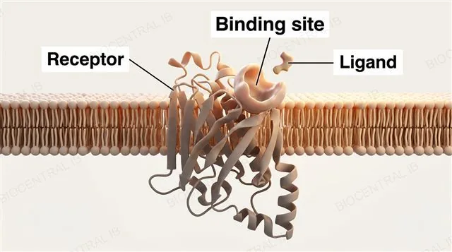
A transmembrane receptor protein spanning a lipid bilayer with a pocket-shaped binding site, a small ligand molecule approaching it, labeled receptor, binding site and ligand
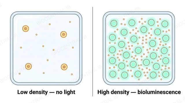
Two panels comparing a low-density bacterial population with no glow to a high-density bacterial population glowing once a signalling-molecule threshold is crossed, labeled low density no light and high density bioluminescence
C2.1.2

When enough bacteria are listening

Quorum sensing is bacterial signalling in which cells secrete and detect a signalling molecule whose concentration reflects local population density, illustrated here by bioluminescence in the marine bacterium Vibrio fischeri.
  • Every cell continuously secretes a small signalling molecule (an autoinducer); at low density this diffuses away and stays too dilute to have any effect on the bacteria around it.
  • Once enough bacteria are packed together, local autoinducer concentration crosses a threshold, binds a receptor, and switches on the genes needed for bioluminescence across the whole population almost simultaneously.
Why it mattersVibrio fischeri lives in some squid species' light organs, where its density-triggered glow camouflages the squid from predators by matching moonlight filtering down from above.
C2.1.3

Four categories, one job

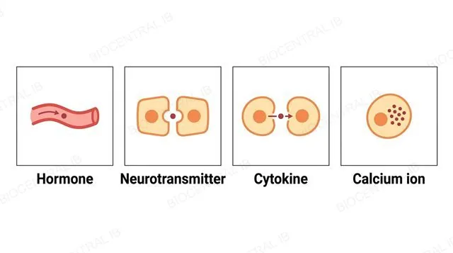
The syllabus groups animal signalling chemicals into four functional categories, distinguished by where they are made, how far they travel, and what they act on.
Hormone
Secreted into the blood and carried, often over the whole body, to distant target cells.
Neurotransmitter
Released by a neuron; diffuses across a narrow synaptic gap to one adjacent cell.
Cytokine
A small signalling protein used mainly for local communication between immune cells.
Calcium ion (Ca²⁺)
Acts as a second messenger entirely inside the cell that produces it.
Why it mattersAn exam question can ask you to identify which category a described signal belongs to from its behaviour alone, not just its name.
Four panels showing the four functional categories of signalling chemical: hormone in the bloodstream, neurotransmitter at a synaptic gap, cytokine between adjacent immune cells, and calcium ion staying inside one cell
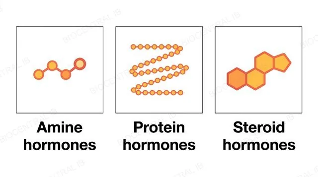
Three panels showing the three chemical groups of hormones as abstract molecular icons, labeled amine hormones, protein hormones and steroid hormones
C2.1.4

Three very different kinds of hormone

Hormones are chemically diverse — amines, proteins and steroids are the three chemical groups named on the syllabus, each suited to a different route into, and effect on, a target cell.
  • Amine hormones (such as epinephrine) and protein hormones (such as insulin) are hydrophilic, so they cannot cross the plasma membrane and instead bind a transmembrane receptor on the cell surface.
  • Steroid hormones (such as oestradiol, progesterone and testosterone) are lipid-derived from cholesterol and hydrophobic, so they can diffuse directly through the membrane to reach a receptor inside the cell.
Why it mattersA hormone's chemical group predicts its receptor type before you even see the mechanism — hydrophilic hormones use surface receptors, hydrophobic ones use intracellular receptors.
C2.1.4

One job, many different molecules

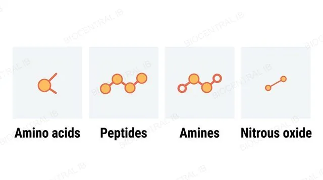
A wide range of chemical substances can act as neurotransmitters, including amino acids, peptides, amines and even the gas nitrous oxide (nitric oxide).
  • Amino acids such as glutamate, peptides, and amines such as acetylcholine and dopamine are all stored in vesicles at the presynaptic membrane and released by exocytosis to act on a surface receptor.
  • Nitrous oxide is the unusual exception: as a small, uncharged gas it diffuses directly through membranes rather than being stored in vesicles or requiring a surface receptor to act.
Why it mattersThis chemical range shows there is no single "neurotransmitter molecule" — what unites these substances is their role at a synapse, not any shared chemical structure.
Four panels showing the four chemical groups that can serve as neurotransmitters as abstract molecular icons, labeled amino acids, peptides, amines and nitrous oxide
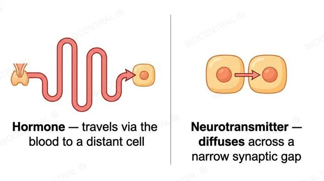
Two panels: a hormone travelling via a long blood-vessel tube to a distant target cell, and a neurotransmitter diffusing across a narrow synaptic gap to an adjacent cell, each with one single-direction arrow
C2.1.5

Reaching one cell, or the whole body

Hormones and neurotransmitters illustrate two very different scales of signalling: distant, body-wide effects and localized, single-cell effects.
Hormone
  • Transported by the blood system to target cells anywhere in the body
  • Reaches its target over seconds to minutes, limited by circulation time
  • One gland can influence many distant tissues at once
Neurotransmitter
  • Diffuses only across a narrow synaptic gap, tens of nanometres wide
  • Reaches its target in a fraction of a millisecond
  • Signals just the one adjacent postsynaptic cell
Why it mattersThis speed-versus-reach trade-off is why the nervous system handles split-second responses while the endocrine system handles slower, longer-lasting, body-wide regulation.
Part two

Receptor types and signal transduction

C2.1.6 – C2.1.9
C2.1.6

Two places a receptor can sit

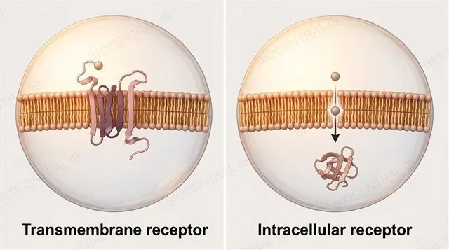
Receptors fall into two structural classes depending on whether their ligand can cross the plasma membrane: transmembrane receptors and intracellular receptors.
Transmembrane
  • Spans the membrane: hydrophobic amino acids sit in the fatty acid core, hydrophilic amino acids face the cytoplasm and extracellular fluid
  • Binds a hydrophilic ligand that cannot cross the membrane, e.g. a protein hormone
Intracellular
  • Sits free in the cytoplasm or nucleus, built almost entirely from hydrophilic amino acids
  • Binds a small, hydrophobic ligand able to diffuse straight through the membrane, e.g. a steroid hormone
Why it mattersAn amino acid's hydrophilic or hydrophobic side chain has to match the chemical environment it sits in — that single principle explains both receptor architectures.
Two panels comparing a transmembrane receptor spanning the membrane with hydrophobic and hydrophilic regions to a separate intracellular receptor in the cytoplasm with a ligand diffusing toward it, labeled transmembrane receptor and intracellular receptor
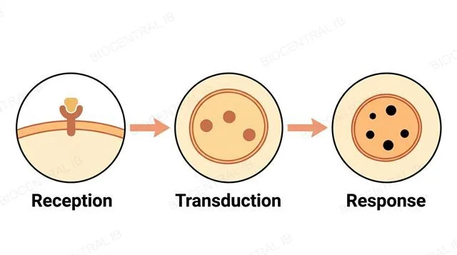
A three-stage flowchart: receptor and ligand docking, then a relay chain inside the cell, then an altered cell response, connected by two single-arrowhead arrows, labeled reception, transduction and response
C2.1.7

One binding event, a chain of responses

Signal transduction is the sequence of responses set off inside a cell once a signalling chemical has bound its receptor, converting an external signal into an internal one.
  • Cell signalling generally proceeds through three stages in order: reception (ligand binds receptor), transduction (a relay of intracellular responses), and response (the cell's eventual altered activity or gene expression).
  • Because each step in the relay can pass the signal to the next molecule, and enzyme-catalysed steps can multiply it, a single receptor-binding event can be converted into a large, amplified response.
Why it mattersThis three-stage framework — reception, transduction, response — is the model to apply to every specific pathway described later in this topic.
C2.1.8

A receptor that is also a channel

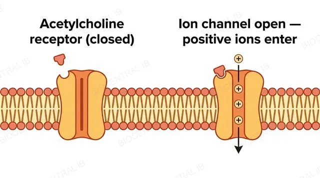
The acetylcholine receptor is a transmembrane receptor built as a ligand-gated ion channel — binding directly opens a channel that is part of the same protein, with no separate messenger needed.
  • When acetylcholine binds, the receptor's channel opens and allows positively charged ions to diffuse into the cell, moving down their electrochemical gradient without requiring any additional energy input.
  • This ion movement changes the voltage across the plasma membrane — a shift that, if large enough, may trigger further changes such as an action potential in the postsynaptic cell.
Why it mattersThis direct, one-step mechanism is faster than any receptor pathway that relies on a second-messenger cascade, which matters at a synapse where timing is critical.
Before and after panels of the acetylcholine receptor: closed channel with ligand outside, then ligand docked with the channel open and three positive ions flowing in through a single-direction arrow
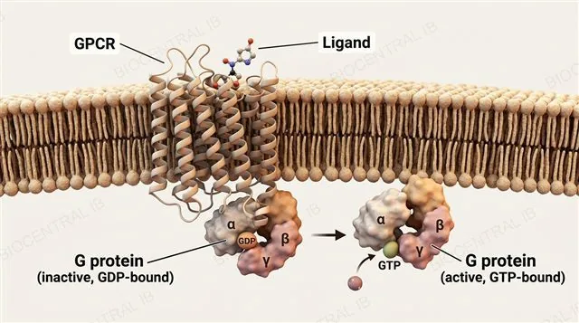
A G protein-coupled receptor spanning the membrane with a docked ligand, and the associated G protein shown before and after activation, labeled GPCR, ligand, G protein inactive GDP-bound and G protein active GTP-bound
C2.1.9

The switch behind an entire receptor family

A G protein-coupled receptor (GPCR) is a transmembrane receptor that, once bound by its ligand, activates an associated G protein to relay the signal onward inside the cell.
  • Ligand binding causes a conformational change in the GPCR, which activates the G protein by promoting exchange of bound GDP (guanosine diphosphate) for GTP (guanosine triphosphate), switching the G protein from inactive to active.
  • Humans express a very large number of distinct GPCRs, each with its own ligand specificity, letting one shared relay mechanism serve an enormous range of different signalling chemicals.
Why it mattersBecause so many receptors share this one mechanism, drugs that target GPCRs make up a substantial share of all medicines currently in clinical use.
Quick check

An acetylcholine receptor and a G protein-coupled receptor are both transmembrane receptors, but they relay a signal very differently. What is the key mechanistic difference?

The acetylcholine receptor opens its own built-in ion channel directly; the GPCR instead activates a separate G protein
The acetylcholine receptor requires cAMP as a second messenger; the GPCR does not
Only the GPCR is a protein — the acetylcholine receptor is a lipid
The acetylcholine receptor works only in plants; GPCRs work only in animals
Correct answer: direct channel vs G protein relay. The acetylcholine receptor is a ligand-gated ion channel — binding opens a channel built into the same protein. A GPCR instead undergoes a conformational change that activates a separate G protein, which goes on to affect a further target enzyme such as adenylyl cyclase.
Part three

Epinephrine and insulin: two transmembrane pathways

C2.1.10 – C2.1.11
C2.1.10

From one hormone to a flood of glucose

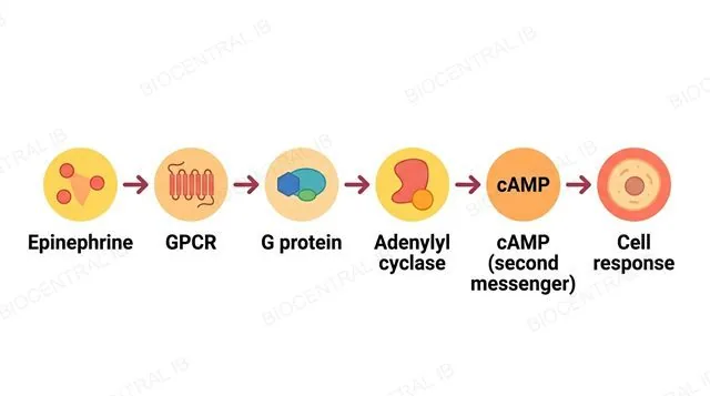
Epinephrine (adrenaline) acts through a G protein-coupled receptor, activating a G protein that in turn activates the enzyme adenylyl cyclase, which converts ATP (adenosine triphosphate) into cyclic AMP (cAMP), the second messenger for this pathway.
  • cAMP goes on to activate a further enzyme cascade inside the cell, ultimately triggering the breakdown of stored glycogen and the release of glucose into the blood.
  • Because each step from adenylyl cyclase onward is catalysed by an enzyme acting on many substrate molecules, the pathway amplifies hugely: a single epinephrine molecule can ultimately release millions of glucose molecules.
Why it mattersThis amplification is why only a tiny concentration of circulating epinephrine is needed to produce a rapid, whole-body rise in blood glucose during a stress response.
A six-step flowchart of the epinephrine signalling cascade connected by five single-arrowhead arrows, labeled epinephrine, GPCR, G protein, adenylyl cyclase, cAMP second messenger and cell response
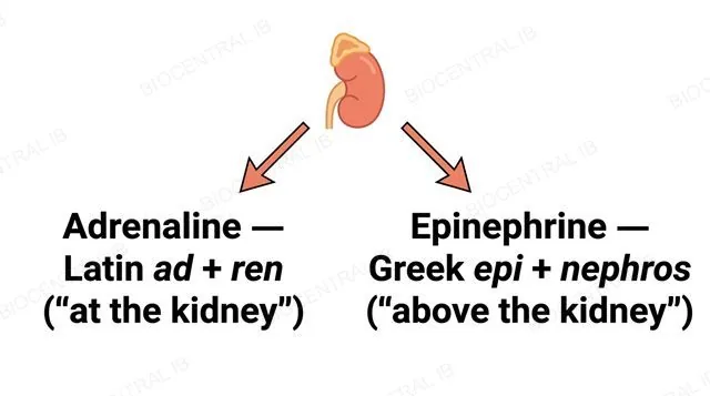
An adrenal-gland icon with two arrows pointing to two text panels showing the Latin origin of adrenaline and the Greek origin of epinephrine, both referring to the same hormone
C2.1.10

One hormone, two names

"Adrenaline" and "epinephrine" name the exact same hormone, and both were coined independently based on its production by the adrenal gland.
  • "Adrenaline" comes from the Latin ad (at) and ren (kidney); "epinephrine" comes from the Greek epi (above) and nephros (kidney) — different languages, describing the same location above the kidney.
  • Both terms have persisted in common use in different parts of the world, rather than one replacing the other, even though they refer to one well-studied molecule.
Why it mattersThis is a Nature of Science point: even settled, well-understood science still depends on international cooperation and convention for something as basic as naming a molecule.
C2.1.11

A receptor that phosphorylates itself

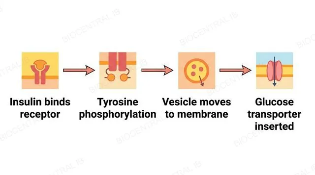
Insulin binds a transmembrane receptor that has its own built-in tyrosine kinase activity — a receptor type distinct from both the acetylcholine receptor and any GPCR.
  • Binding of insulin to its receptor causes phosphorylation of tyrosine amino acids inside the cell, setting off a sequence of further reactions without any G protein or ion channel involved.
  • This sequence of reactions ends with vesicles containing glucose transporter proteins moving to, and fusing with, the plasma membrane, increasing the cell's capacity to take up glucose from the blood.
Why it mattersInsulin and epinephrine both signal through transmembrane receptors, but their distinct mechanisms — tyrosine kinase versus GPCR/cAMP — produce opposite effects on blood glucose.
A four-step flowchart of the insulin tyrosine kinase receptor pathway connected by three single-arrowhead arrows, labeled insulin binds receptor, tyrosine phosphorylation, vesicle moves to membrane and glucose transporter inserted
Quick check

In the epinephrine signalling pathway, why does adding an inhibitor of phosphodiesterase (the enzyme that breaks cAMP down to AMP) prolong the cell's response, even after epinephrine itself is removed?

cAMP is not degraded, so it persists and keeps the downstream response active for longer
The G protein becomes permanently bound to epinephrine
Adenylyl cyclase stops producing cAMP altogether
The receptor is destroyed, so no further signal is needed
Correct answer: cAMP persists. Phosphodiesterase normally breaks cAMP down to AMP, terminating the signal. If it is inhibited, cAMP cannot be degraded, so it remains elevated and the downstream response (such as glycogen breakdown) continues even without more epinephrine binding.
Part four

Steroid hormones and feedback regulation

C2.1.12 – C2.1.14
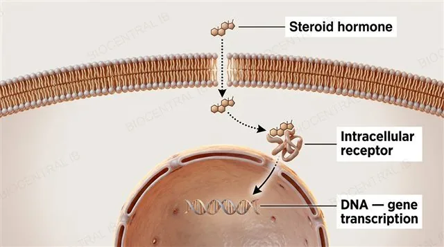
A steroid hormone crossing the plasma membrane, binding an intracellular receptor, and the hormone-receptor complex entering the nucleus to bind DNA, labeled steroid hormone, intracellular receptor and DNA gene transcription
C2.1.12

A hormone that becomes a transcription factor

The steroid hormones oestradiol, progesterone and testosterone act through intracellular receptors that, once activated, bind DNA directly to promote transcription of specific genes.
  • The hormone diffuses through the plasma membrane and binds a site on its receptor in the cytoplasm or nucleus, activating the receptor by causing a conformational change.
  • The activated hormone-receptor complex then binds specific DNA sequences, acting as a transcription factor that promotes transcription of the genes it targets — with no second messenger or ion channel step involved.
Why it mattersBecause new gene transcription, and often new protein synthesis, must happen first, intracellular-receptor pathways are typically slower to start than membrane-receptor pathways, but longer-lasting.
C2.1.13

One hormone, one specified target

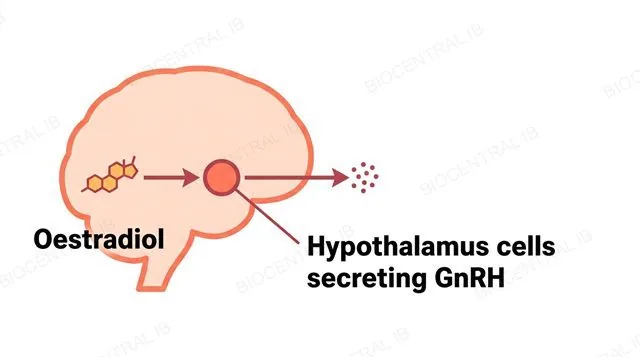
The syllabus limits oestradiol's required effect to cells in the hypothalamus that secrete gonadotropin-releasing hormone (GnRH), a hormone that itself controls further hormone release from the pituitary gland.
  • Oestradiol diffuses into these hypothalamus cells and binds its intracellular receptor, and the activated receptor-hormone complex alters transcription of genes that shape the pattern of GnRH release.
  • Because GnRH in turn governs release of the hormones that regulate the menstrual cycle, oestradiol's action on this one small group of cells has effects reaching well beyond the hypothalamus itself.
Why it mattersThis is the general intracellular-receptor mechanism from the previous slide, applied here to one specific, syllabus-named target cell type.
A simplified brain outline with the hypothalamus highlighted, oestradiol binding that region with a single-direction arrow, labeled oestradiol and hypothalamus cells secreting GnRH
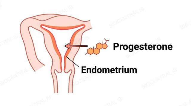
A simplified uterus cross-section with the endometrium highlighted, progesterone binding that lining with a single-direction arrow, labeled progesterone and endometrium
C2.1.13

A second named target, a different tissue

The syllabus limits progesterone's required effect to cells in the endometrium, the lining of the uterus, where its receptor-hormone complex promotes transcription of specific genes.
  • Progesterone diffuses into endometrial cells and binds its intracellular receptor there, and the activated complex promotes transcription of genes that help maintain and prepare the endometrium for implantation.
  • Oestradiol and progesterone act through the identical general mechanism described in C2.1.12 — the difference between them is entirely which target cell type each one is specified to act on.
Why it mattersDo not swap these two hormone-target pairs: oestradiol is paired with GnRH-secreting hypothalamus cells, progesterone with endometrial cells.
C2.1.14

Two ways a pathway can regulate itself

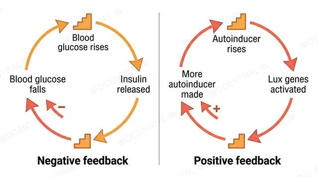
Cell signalling pathways can be regulated by negative feedback, which stabilises a variable, or positive feedback, which reinforces it — the syllabus limits this to understanding the difference and one example of each.
Negative feedback
  • Reduces or reverses the stimulus, restoring a stable set point
  • Rising blood glucose → insulin released → blood glucose falls → less insulin released
Positive feedback
  • Reinforces the stimulus, driving the system further from its starting state
  • Lux genes activated → more autoinducer made → even more activation
Why it mattersMost homeostatic regulation in the body relies on negative feedback; positive feedback is reserved for the rarer cases where a fast, self-reinforcing switch is the useful outcome.
Two circular loop diagrams: negative feedback for blood glucose and insulin with an inward minus arrow, and positive feedback for quorum sensing autoinducer with an inward plus arrow, labeled negative feedback and positive feedback

Key vocabulary

Worth being able to define in a single sentence each

Ligand
The IB term for any signalling chemical that binds specifically to a receptor.
Signal transduction
The sequence of responses set off inside a cell once a ligand has bound its receptor.
Second messenger
A small intracellular molecule, such as cAMP, that relays a signal onward once a receptor has been activated.
GPCR
A G protein-coupled receptor: a transmembrane receptor that activates an associated G protein once bound by its ligand.
Quorum sensing
Bacterial signalling in which gene expression changes once population density crosses a threshold.
Tyrosine kinase receptor
A transmembrane receptor, such as the insulin receptor, that phosphorylates tyrosine residues once activated.
Intracellular receptor
A receptor in the cytoplasm or nucleus, binding a ligand small and hydrophobic enough to cross the membrane.
Positive feedback
Regulation in which a pathway's output reinforces the original stimulus.
Negative feedback
Regulation in which a pathway's output reduces or reverses the original stimulus.

Where this shows up again

B1.2
Many hormones are proteins (B1.2). Using insulin as an example, explain how the primary and tertiary structure of a protein hormone enables specific binding to its receptor.
C1.2
Signalling pathways often involve phosphorylation of target proteins. Explain how ATP from respiration (C1.2) provides the phosphate groups used in receptor tyrosine kinase signalling.
C2.2
Neurotransmitters such as acetylcholine act as ligands in the nervous system (C2.2). Compare the speed and mechanism of neurotransmitter signalling with that of endocrine hormone signalling.
D2.2
Gene expression (D2.2) is a common endpoint of signal transduction. Explain how a steroid hormone-receptor complex can directly activate transcription of specific genes.

C2.1 Chemical signalling — one-page recap

Screenshot this slide to revise from

Receptors & ligands
  • Receptor: protein with a binding site complementary to a specific ligand; binding causes a conformational change.
  • Quorum sensing: Vibrio fischeri bioluminescence switches on once autoinducer crosses a density threshold.
Categories & chemistry
  • Four functional categories: hormone, neurotransmitter, cytokine, calcium ion (Ca²⁺) — differ in range and role.
  • Hormones: amine, protein, steroid. Neurotransmitters: amino acid, peptide, amine, nitrous oxide.
Distance & receptor location
  • Hormones travel via blood to distant cells; neurotransmitters diffuse across a synaptic gap.
  • Transmembrane receptors bind hydrophilic ligands; intracellular receptors bind hydrophobic ligands that cross the membrane.
Transduction & two receptor types
  • Reception → transduction → response. Acetylcholine receptor: ligand-gated ion channel, changes membrane potential directly.
  • GPCR: activates a G protein (GDP→GTP exchange) to relay the signal onward.
Epinephrine & insulin
  • Epinephrine: GPCR → G protein → adenylyl cyclase → cAMP second messenger → glycogen breakdown, amplified.
  • Insulin: tyrosine kinase receptor → phosphorylation → glucose transporter vesicles move to the membrane.
Steroids & feedback
  • Intracellular receptors (oestradiol → hypothalamus/GnRH; progesterone → endometrium) bind DNA to promote transcription.
  • Negative feedback stabilises (insulin/glucose); positive feedback reinforces (quorum sensing).

Small signal, cell-wide change

From a single ligand docking into a single binding site to an entire gene switched on or a whole pathway amplified a millionfold, every mechanism in this lesson comes down to one idea: a receptor converts a chemical message into a cellular decision.
C2.1 Chemical signalling · BioCentral IB
Use ↓ ↑ or click to navigate
01 / 20