BioCentral IBBioCentral IB
IB Biology · Theme C · C2.2

Neural signalling

From a single sodium ion crossing a membrane to billions of neurons firing together, this lesson traces how the nervous system generates, propagates, and passes on an electrical signal — and where synapses turn that signal chemical again.
Guiding questions

How are electrical signals generated and moved within neurons?

How can neurons interact with other cells?

Part one

Neuron structure and the resting potential

C2.2.1 – C2.2.2
C2.2.1

One cell body, many fibres

A neuron is a specialized cell of the nervous system whose cytoplasm and nucleus form a cell body, with elongated nerve fibres of varying length projecting outward to conduct electrical impulses.
  • A single long fibre called the axon carries impulses away from the cell body, often over a considerable distance to a target cell.
  • Multiple shorter fibres called dendrites project from the cell body and typically receive incoming signals from other neurons.
  • Both the axon and dendrites conduct electrical impulses — it is their number and length that distinguishes one from the other, not signalling direction alone.
Why it mattersThe IB data booklet provides a labelled diagram of a generalized neuron — learn to identify the cell body, axon and dendrites on sight, since exam questions often ask you to interpret this exact figure.
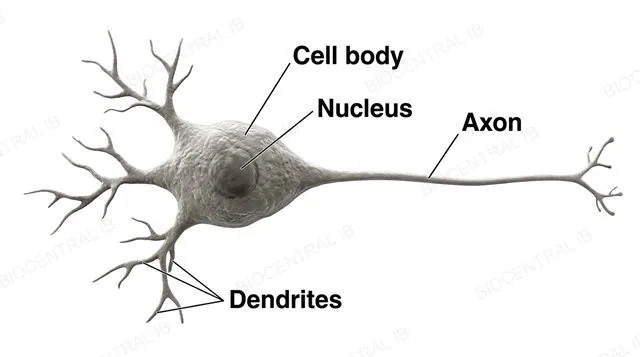
A generalized neuron showing the cell body with nucleus, one long axon, and multiple shorter dendrites projecting from the cell body, labeled cell body, nucleus, axon and dendrites
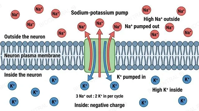
A neuron membrane cross-section showing the sodium-potassium pump moving three sodium ions out of the cell and two potassium ions into the cell, with concentration labels higher sodium outside and higher potassium inside, labeled sodium-potassium pump, Na+ out and K+ in
C2.2.2

Charging the membrane at rest

The resting potential is the voltage difference across a neuron's plasma membrane when it is not conducting an impulse, generated and maintained by ATP-driven pumping of sodium and potassium ions in opposite directions.
  • The sodium-potassium pump uses energy from ATP to actively transport sodium ions out of the neuron and potassium ions into the neuron, against their concentration gradients.
  • Because this pumping leaves relatively more positive ions outside the membrane than inside, the membrane becomes polarized — a voltage difference exists across it.
  • The resulting membrane potential is negative, roughly −70 mV, with the inside of a resting neuron negative relative to the outside.
Why it mattersThis pumping never stops, even at rest — a neuron continuously spends ATP just to maintain the gradients that make it ready to fire an impulse at any moment.
Part two

Action potentials and impulse speed

C2.2.3 – C2.2.4
C2.2.3

An impulse is an electrical event

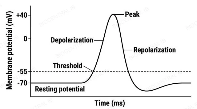
A nerve impulse, or action potential, is a rapid, temporary reversal of the membrane potential that is generated and then propagated along a nerve fibre — it is electrical because it involves movement of positively charged ions.
  • An action potential only fires once a stimulus depolarizes the membrane to a threshold potential; weaker, sub-threshold stimuli produce no action potential at all.
  • Once threshold is reached, the action potential fires as an all-or-nothing event — every action potential in a given neuron reaches the same peak size, regardless of the stimulus.
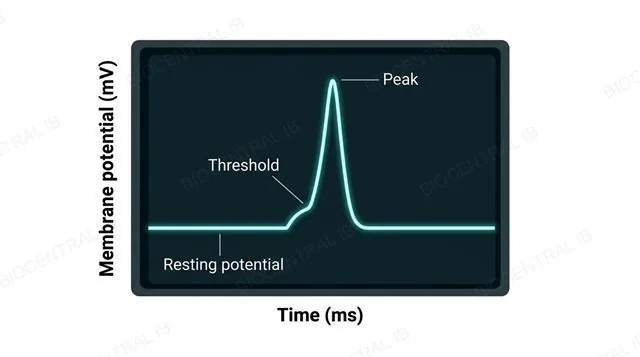
  • An oscilloscope trace of a stimulated axon can record this rapid rise and fall in membrane potential over time.
Why it mattersAll-or-nothing firing means stimulus strength can't be coded in one action potential's size — it must be coded some other way, covered later in this lesson.
A voltage against time graph of a single action potential showing resting potential, threshold, rapid depolarization to a positive peak, and repolarization back down, labeled resting potential, threshold and peak
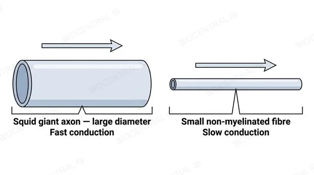
Two panels comparing a large-diameter squid giant axon conducting an impulse quickly with a small-diameter non-myelinated fibre conducting the same impulse slowly, each with one directional arrow along the axon, labeled squid giant axon large diameter fast and small non-myelinated fibre slow
C2.2.4

Bigger axons carry impulses faster

Conduction velocity, the speed at which an impulse travels along a fibre, varies considerably between neurons and depends strongly on axon diameter.
  • The squid giant axon, with an unusually large diameter, conducts impulses far faster than the small, non-myelinated nerve fibres typical of many other invertebrates.
  • A wider axon offers less internal resistance to the local current that drives propagation, so an impulse spreads along it more quickly even without any insulation.
  • This diameter effect is a genuinely separate factor from myelination, covered later in this lesson, which achieves fast conduction by a completely different route.
Why it mattersThe squid giant axon is a classic experimental model precisely because its large size makes it physically easy to record from directly.
C2.2.4

Reading the strength of a relationship

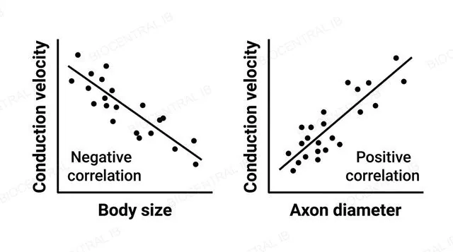
Conduction velocity is negatively correlated with animal body size but positively correlated with axon diameter — a required application-of-skills point for interpreting data like this.
  • A negative correlation means one variable decreases as the other increases; a positive correlation means both variables increase together — correlation coefficients quantify the strength of each relationship.
  • The coefficient of determination, R², expresses the proportion of variation in conduction velocity that is explained by variation in the independent variable.
  • Correlation alone does not establish that one variable directly causes changes in the other, only that a statistical relationship exists in the data.
Why it mattersExpect a calculation or interpretation question that hands you a correlation coefficient or R² value directly and asks what it means about the strength of a stated relationship.
Two small scatter graphs side by side: one showing conduction velocity plotted against body size with a downward-sloping trend line for a negative correlation, and one showing conduction velocity plotted against axon diameter with an upward-sloping trend line for a positive correlation, labeled negative correlation body size and positive correlation axon diameter
Part three

Synapses and neurotransmission

C2.2.5 – C2.2.7
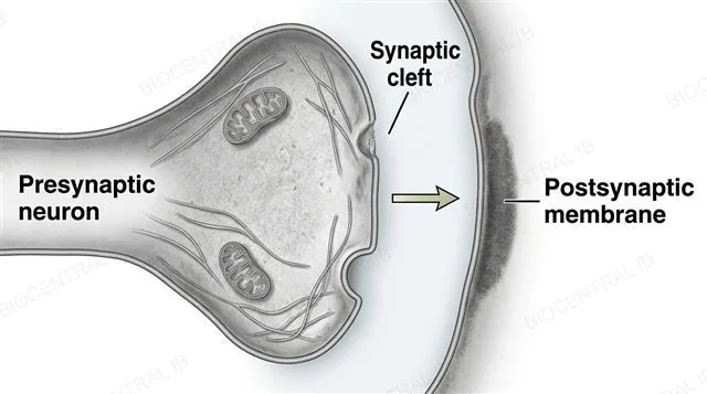
A chemical synapse showing a presynaptic terminal, a narrow synaptic cleft, and a postsynaptic membrane, with a single arrow pointing from the presynaptic terminal across the cleft to the postsynaptic membrane, labeled presynaptic neuron, synaptic cleft and postsynaptic membrane
C2.2.5

A junction with only one way through

A synapse is a junction between two neurons, or between a neuron and an effector cell, across which a signal passes — this course limits synapses to chemical synapses only.
  • At a typical chemical synapse, the presynaptic cell releases a chemical signal that the postsynaptic cell detects, so information can only flow in one direction, presynaptic to postsynaptic.
  • Synapses aren't limited to connections between two neurons — the neuromuscular junction, between a motor neuron and a muscle fibre, is also a synapse under this definition.
  • Electrical synapses exist in some organisms but are excluded from this course; every synapse discussed here is a chemical one.
Why it mattersUnidirectional signalling at synapses is what gives a neural circuit its fixed direction of information flow, from receptor cells through to an effector.
C2.2.6

Calcium turns an impulse chemical

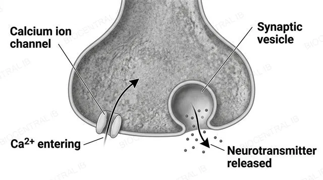
Release of neurotransmitter from the presynaptic membrane is triggered by depolarization of that membrane, which causes uptake of calcium ions that act as an internal signalling chemical inside the neuron.
  • When an action potential arrives at the presynaptic terminal, the resulting depolarization opens channels that let calcium ions enter the presynaptic cell from outside.
  • This calcium influx signals synaptic vesicles, each loaded with neurotransmitter, to fuse with the presynaptic membrane and release their contents into the synaptic cleft.
  • Without calcium entry, depolarization of the presynaptic membrane alone is not sufficient to trigger neurotransmitter release.
Why it mattersCalcium acting as an internal signal, rather than the neurotransmitter itself, is the crucial link that converts an electrical impulse into a chemical one at every synapse.
A presynaptic terminal showing a calcium ion channel with calcium ions entering the cell through it, and a synaptic vesicle fusing with the presynaptic membrane to release neurotransmitter molecules into the synaptic cleft, labeled calcium ion channel, Ca2+ entering, synaptic vesicle and neurotransmitter released
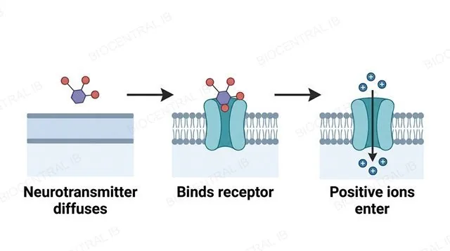
A three-step flowchart showing a neurotransmitter diffusing across the synaptic cleft, then binding a transmembrane receptor on the postsynaptic membrane, then positive ions entering through an opened channel, connected by two single-arrowhead arrows, labeled neurotransmitter diffuses, binds receptor and positive ions enter
C2.2.7

One neurotransmitter, many synapses

An excitatory postsynaptic potential (EPSP) is generated when neurotransmitter diffuses across the synaptic cleft and binds transmembrane receptors on the postsynaptic membrane, opening ion channels and depolarizing that membrane toward threshold.
  • Acetylcholine is the syllabus example of a neurotransmitter that generates an EPSP, and it's found in many types of synapse, including the neuromuscular junction.
  • Binding of acetylcholine to its transmembrane receptor lets positively charged ions cross the postsynaptic membrane, moving its potential closer to the threshold needed for an action potential.
  • An EPSP is a graded change in potential, not an all-or-nothing event — only the resulting action potential, if threshold is reached, is all-or-nothing.
Why it mattersThe receptor sits on the postsynaptic side, not the presynaptic side — a common point of confusion worth fixing early.
Quick check

A stimulus depolarizes a neuron's membrane, but not enough to reach the threshold potential. What happens?

No action potential is generated — the stimulus produces only a small, local change in potential
A smaller-than-normal action potential fires
The neuron fires an action potential at half its normal speed
The resting potential becomes permanently positive
Correct answer: no action potential at all. Action potentials are all-or-nothing — threshold must be reached for voltage-gated sodium channels to open and trigger the full event. A sub-threshold stimulus produces only a local, non-propagated change in membrane potential, never a smaller impulse.
Part four

The action potential mechanism · HL

C2.2.8 – C2.2.11
C2.2.8 · HL

Two channels, opposite directions

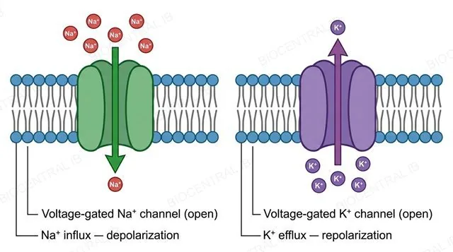
Depolarization and repolarization during an action potential are produced by voltage-gated sodium and potassium channels opening in sequence, once the membrane reaches its threshold potential.
  • When the membrane depolarizes to threshold, voltage-gated sodium channels open, and sodium ions rush into the neuron down their electrochemical gradient, driving the rapid depolarization phase.
  • At the peak of the action potential, voltage-gated sodium channels inactivate while voltage-gated potassium channels open, so potassium ions flow out of the neuron, repolarizing the membrane toward resting potential.
  • Reaching the threshold potential in the first place is essential — below it, voltage-gated sodium channels simply do not open.
Why it mattersThe sodium-potassium pump plays no direct role in this rapid cycle — its slower job is restoring the ion gradients afterwards, not causing the voltage change itself.
Two panels of a neuron membrane at the threshold potential: the first showing an open voltage-gated sodium channel with sodium ions moving into the cell, the second showing an open voltage-gated potassium channel with potassium ions moving out of the cell, each with one single-direction arrow, labeled voltage-gated sodium channel open Na+ influx depolarization and voltage-gated potassium channel open K+ efflux repolarization
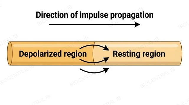
A length of unmyelinated axon showing one depolarized region and an adjacent resting region, with short arrows showing sodium ions diffusing from the depolarized region toward the resting region both inside and outside the axon, plus one larger arrow showing the single direction of impulse propagation along the axon, labeled depolarized region, resting region and direction of impulse propagation
C2.2.9 · HL

How an impulse travels without moving

An action potential is propagated along a nerve fibre because local currents — diffusion of sodium ions inside and outside the axon — bring the adjacent membrane to threshold.
  • Sodium ions that entered during depolarization diffuse locally to the neighbouring, still-resting region of membrane, both inside and just outside the axon.
  • This local ion movement raises the neighbouring membrane's potential to threshold, opening voltage-gated sodium channels there and regenerating a full action potential at that new position.
  • Because the region just behind the impulse is briefly refractory and unable to fire again, this regeneration moves in one direction only, away from where it began.
Why it mattersThe action potential is not a signal passively flowing down a wire — it is actively regenerated, in full, at every point along an unmyelinated fibre.
C2.2.10 · HL

Watching a membrane's voltage change

An oscilloscope displays membrane potential against time, letting both the resting potential and the depolarization and repolarization phases of an action potential be identified and interpreted directly from the trace.
  • The flat, unchanging portion of a trace before a stimulus arrives corresponds to the resting potential; a sharp upward deflection marks the start of depolarization.
  • The number of impulses recorded within a known time window can be measured and converted to a frequency, in hertz, using impulses divided by time in seconds.
  • Comparing the shape, size and timing of traces from different neurons or conditions links a trace back to real, specific cellular events.
Why it mattersReading an oscilloscope trace is an application-of-skills task in its own right — expect to identify a labelled feature or calculate a frequency from a given trace.
An oscilloscope screen displaying a trace of membrane potential against time, with a flat resting-potential section followed by a sharp action-potential spike, axes labeled membrane potential millivolts and time milliseconds, and the trace itself labeled resting potential, threshold and peak
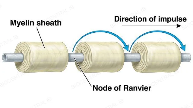
A myelinated axon with several nodes of Ranvier as gaps in the myelin sheath, with arrows showing the action potential jumping from one node directly to the next in a single direction along the axon, labeled myelin sheath, node of Ranvier and direction of impulse
C2.2.11 · HL

Jumping from node to node

Saltatory conduction is the faster form of impulse propagation in myelinated fibres, where ion pumps and channels are clustered at gaps in the myelin sheath called nodes of Ranvier.
  • The myelin sheath insulates long stretches of the axon between nodes, so the membrane beneath it cannot generate an action potential directly.
  • Because ion channels are concentrated at the nodes of Ranvier, the action potential is only regenerated at these points, and appears to jump from node to node along the fibre.
  • Since far fewer points need active regeneration compared with a continuously conducting unmyelinated fibre, saltatory conduction achieves substantially faster impulses for a similar axon diameter.
Why it mattersMyelin itself does not conduct the signal — it's a passive insulator that forces conduction to become node-to-node instead of continuous.
Part five

Synaptic transmission in depth · HL

C2.2.12 – C2.2.14
C2.2.12 · HL

Blocking the receiving end

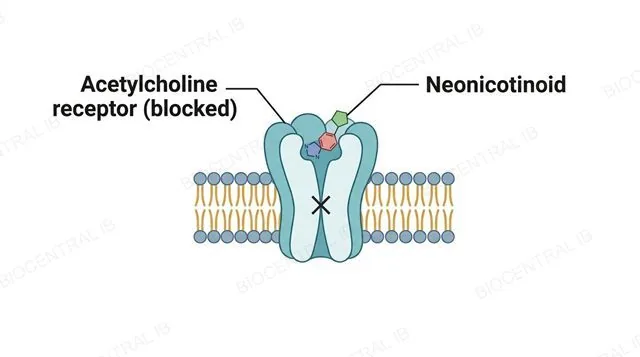
Neonicotinoid pesticides are the syllabus example of an exogenous chemical that blocks synaptic transmission by acting on the postsynaptic membrane, rather than the presynaptic terminal.
  • Neonicotinoids bind to acetylcholine receptors on the postsynaptic membrane, occupying the site that acetylcholine itself would normally bind.
  • Because the receptor is blocked rather than activated normally, this binding disrupts the insect's usual synaptic transmission at cholinergic synapses, ultimately causing paralysis and death.
  • This receptor-blocking mechanism is distinct from an enzyme-blocking mechanism — neonicotinoids do not act on any enzyme that breaks down neurotransmitter.
Why it mattersNeonicotinoids are widely used agricultural insecticides, which has made their effect on non-target pollinating insects a genuine environmental concern.
A postsynaptic membrane with an acetylcholine receptor occupied by a neonicotinoid molecule instead of acetylcholine, with a crossed-out X symbol showing the channel stays closed, labeled acetylcholine receptor blocked and neonicotinoid
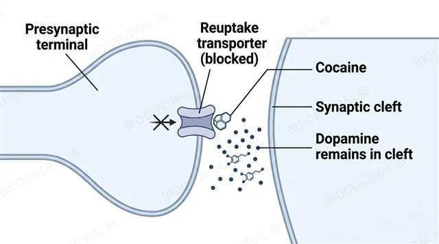
A presynaptic terminal with a reuptake transporter protein blocked by a cocaine molecule, and dopamine molecules accumulating in the synaptic cleft with a crossed-out arrow showing blocked reuptake, labeled reuptake transporter blocked, cocaine and dopamine remains in cleft
C2.2.12 · HL

Blocking the sending end

Cocaine is the syllabus example of a drug that affects synaptic transmission by blocking reuptake of neurotransmitter at the presynaptic terminal, rather than acting at the postsynaptic receptor.
  • Normally, a transporter protein in the presynaptic membrane removes neurotransmitter such as dopamine from the synaptic cleft after it has acted, ending the signal and recycling the neurotransmitter.
  • Cocaine blocks this reuptake transporter, so neurotransmitter stays in the synaptic cleft for longer, prolonging its stimulatory effect on the postsynaptic membrane.
  • Because neonicotinoids and cocaine act at opposite sides of the synapse and by different mechanisms, they should never be treated as interchangeable examples.
Why it mattersCocaine's prolonged dopamine signalling in reward pathways of the brain is central to why the drug is so strongly associated with addiction.
C2.2.13 · HL

Pushing a membrane further from firing

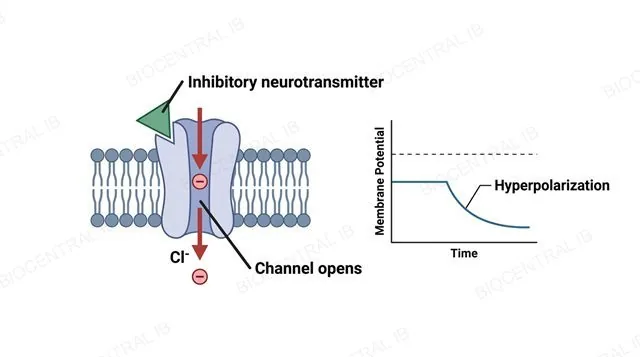
An inhibitory postsynaptic potential (IPSP) is generated when an inhibitory neurotransmitter binds its receptor and causes the postsynaptic membrane to become hyperpolarized — more negative than its resting potential.
  • Hyperpolarization moves the membrane potential further from the threshold needed to trigger an action potential, making the postsynaptic neuron less likely to fire.
  • An IPSP is a real, measurable change in potential, not simply the absence of excitatory input — it actively works against any EPSPs arriving at the same time.
  • Inhibitory and excitatory neurotransmitters can act at entirely different synapses onto the very same postsynaptic neuron, giving it competing inputs to reconcile.
Why it mattersMixing up EPSP and IPSP — depolarizing toward threshold versus hyperpolarizing away from it — is one of the most common exam errors on this topic.
A postsynaptic membrane with an inhibitory neurotransmitter binding its receptor and a channel opening, with a small inset graph showing the membrane potential moving downward away from the threshold line, labeled inhibitory neurotransmitter, channel opens and hyperpolarization
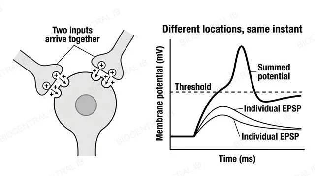
A postsynaptic neuron receiving EPSP inputs from two different presynaptic neurons arriving at the same instant, with a graph showing two small individual EPSP curves and one larger bold summed-potential curve crossing the dashed threshold line, labeled two inputs arrive together, individual EPSP, summed potential and threshold
C2.2.14 · HL

Adding up different inputs

Spatial summation is the addition of postsynaptic potentials arriving at the same moment from different synapses on a postsynaptic neuron.
  • Two or more presynaptic neurons can each contribute an EPSP arriving at essentially the same instant, from different points on the postsynaptic neuron's surface.
  • Each individual EPSP may be too small to reach threshold alone, but their combined, summed effect at that instant can trigger an action potential.
  • The name reflects where the inputs come from — different locations — rather than when they arrive, which is what temporal summation is about instead.
Why it mattersA single synapse rarely decides whether a neuron fires — spatial summation lets it integrate simultaneous input from many sources at once.
C2.2.14 · HL

Adding up the same input, over time

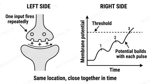
Temporal summation is the addition of postsynaptic potentials generated in quick succession by a single presynaptic neuron at one synapse.
  • If one presynaptic neuron fires repeatedly fast enough, each new EPSP arrives before the last one has fully decayed, so their effects build on top of each other.
  • This build-up can push the membrane potential up to threshold even though no single impulse from that synapse would have been enough alone.
  • The same location is involved each time — only the timing of repeated inputs changes, which is what distinguishes it from spatial summation.
Why it mattersTemporal summation means firing rate, not just whether a synapse fires at all, carries real information the postsynaptic neuron can use.
A postsynaptic neuron receiving three excitatory signals in quick succession from a single presynaptic neuron at one synapse, with a graph showing a staircase-like curve building up over three numbered steps to cross the dashed threshold line, labeled one input fires repeatedly, threshold and potential builds with each pulse
Quick check · HL

A postsynaptic neuron receives an EPSP that would reach threshold on its own. At the same moment, it also receives an IPSP from a different presynaptic neuron. What is the most likely outcome?

The neuron may not fire, because the IPSP's hyperpolarization can counteract the EPSP's depolarization when the two are summed
The neuron always fires, because at least one EPSP reached threshold
The IPSP is ignored entirely, since EPSPs always take priority
The neuron fires twice, once for each signal
Correct answer: summation can prevent firing. Postsynaptic potentials are graded and summate — a simultaneous IPSP hyperpolarizes the membrane, counteracting the EPSP's depolarization. Whether the neuron fires depends on the combined, summed potential at the axon hillock reaching threshold, not on any single input in isolation.
Part six

Pain and consciousness · HL

C2.2.15 – C2.2.16
C2.2.15 · HL

Free endings that sense danger

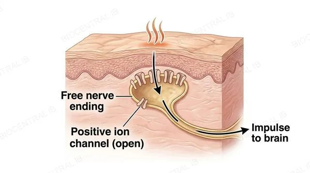
Free nerve endings in the skin detect pain-associated stimuli such as high temperature, acid, or capsaicin, using channels for positively charged ions that open in response.
  • When a stimulus such as high temperature or acid is detected, channels for positively charged ions open in the free nerve ending, allowing these ions to enter.
  • Entry of positive ions brings the local membrane to threshold, generating nerve impulses that pass through the sensory neuron toward the brain.
  • Capsaicin, the compound behind the heat of chilli peppers, opens these same channels directly, which is why it produces a genuine sensation of heat and pain without real tissue-damaging temperature.
Why it mattersPain itself is perceived in the brain, not at the skin — the free nerve ending only detects the stimulus and generates the impulse that eventually reaches it.
A cross-section of skin showing a free nerve ending with open channels for positively charged ions responding to a heat source above it, with positive ions entering the nerve ending and one arrow showing the impulse travelling away toward the brain, labeled free nerve ending, positive ion channel open and impulse to brain
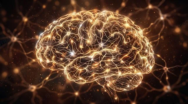
An abstract, warmly lit network of many interconnected glowing neurons forming a brain-like web, with no text or labels, suggesting a single emergent property arising from countless individual connections
C2.2.16 · HL

A property of the whole network

Consciousness is described in this course as an emergent property — one that arises from the interaction of many individual neurons in the brain, and that no single neuron possesses on its own.
  • A single neuron studied in isolation shows no sign of consciousness — the property only appears at the level of a large, interconnected network.
  • This is one example of a wider theme in biology: emergent properties are features of a whole system that cannot be predicted from studying its parts separately.
  • Billions of neurons, each following the same resting-potential, action-potential and synaptic rules covered in this lesson, together give rise to this one emergent property.
Why it mattersThis idea ties the lesson together — every mechanism here, from a single ion pump to a single synapse, scales up into something new once enough of them interact.

Key vocabulary

Worth being able to define in a single sentence each

Resting potential
The voltage difference across a neuron's membrane at rest, generated by active pumping of Na⁺ and K⁺ in opposite directions.
Action potential
An all-or-nothing electrical impulse triggered once a stimulus depolarizes the membrane to threshold.
Synapse
A chemical junction between two neurons, or between a neuron and an effector cell, across which a signal passes in one direction.
Threshold potential
The minimum depolarization needed to trigger an action potential.
EPSP
Excitatory postsynaptic potential: a depolarizing, graded shift in postsynaptic membrane potential toward threshold.
Saltatory conductionHL
The "jumping" of an action potential from node to node in a myelinated fibre, achieving faster impulses.
Refractory periodHL
The brief period after an action potential during which a neuron cannot (absolute) or is less able (relative) to fire again.
IPSPHL
Inhibitory postsynaptic potential: a hyperpolarizing, graded shift in postsynaptic membrane potential away from threshold.
SummationHL
Combining multiple EPSPs and IPSPs at a postsynaptic neuron into one all-or-nothing firing decision.

Where this shows up again

B2.1
The sodium-potassium pump (B2.1) maintains the Na⁺ and K⁺ gradients essential for action potentials. Calculate the number of Na⁺ ions pumped out per ATP if the resting potential is to be maintained.
B1.2
The structure of proteins (B1.2) determines the specificity of neurotransmitter receptors. Explain how the tertiary structure of a ligand-gated ion channel enables it to open only in response to a specific neurotransmitter.
B3.3
At the neuromuscular junction (B3.3), acetylcholine triggers muscle contraction. Compare the postsynaptic response at the NMJ with that at a neuron-neuron synapse in the central nervous system.
C2.1
Many psychoactive drugs affect synaptic transmission. Using cocaine (dopamine reuptake inhibitor) as an example, explain how altering synaptic transmission can lead to addiction.

C2.2 Neural signalling — one-page recap

Screenshot this slide to revise from

Neuron structure & resting potential
  • Neuron: cell body (cytoplasm + nucleus) with one long axon and multiple shorter dendrites, all conducting impulses.
  • Resting potential (~−70 mV, inside negative): maintained by ATP-driven pumping of Na⁺ out and K⁺ in, in opposite directions.
Action potentials & impulse speed
  • Action potential: all-or-nothing electrical event, triggered only once threshold is reached; read on oscilloscope traces.
  • Conduction velocity is negatively correlated with body size, positively correlated with axon diameter (R² quantifies each relationship's strength).
Synapses & neurotransmission
  • Synapse: junction (neuron–neuron or neuron–effector) where a signal passes one direction only; depolarization → Ca²⁺ entry → neurotransmitter release.
  • EPSP: neurotransmitter (e.g. acetylcholine) diffuses across the cleft, binds a receptor, depolarizes the postsynaptic membrane toward threshold.
HL · The action potential mechanism
  • Depolarization = Na⁺ influx via voltage-gated Na⁺ channels; repolarization = K⁺ efflux via voltage-gated K⁺ channels.
  • Local currents regenerate the impulse point-by-point (unmyelinated); saltatory conduction jumps node-to-node at nodes of Ranvier (myelinated) for speed.
HL · Synaptic transmission in depth
  • Neonicotinoids block the postsynaptic acetylcholine receptor; cocaine blocks presynaptic reuptake of neurotransmitter (e.g. dopamine) — different sides, different mechanisms.
  • IPSP hyperpolarizes the postsynaptic membrane; summation (spatial + temporal) combines many EPSPs/IPSPs into one all-or-nothing firing decision.
HL · Pain & consciousness
  • Free nerve endings open positive-ion channels in response to heat, acid or capsaicin, sending impulses to the brain, where pain is perceived.
  • Consciousness is an emergent property arising from the interaction of billions of individual neurons, not a property any single neuron has alone.

Every thought, one ion at a time

From a single sodium ion crossing a membrane to billions of neurons interacting to produce consciousness itself, this lesson traces the nervous system's electrical and chemical signalling from the smallest scale to the largest.
C2.2 Neural signalling · BioCentral IB
Use ↓ ↑ or click to navigate
01 / 20