BioCentral IBBioCentral IB
IB Biology · Theme C · C3.2

Defence against disease

From a chemical shield on skin to a phagocyte's silent hunt and an antibody built to remember, this lesson follows the body's defences from first contact through the adaptive immune response — and the public-health strategies, from vaccines to herd immunity, that turn one person's immunity into a population's protection.
Guiding questions

How do body systems recognize pathogens and fight infections?

What factors influence the incidence of disease in populations?

Part one

First-line and innate defence

C3.2.1 – C3.2.5
C3.2.1

Not every microbe is an enemy

A pathogen is a disease-causing organism; the term usually covers viruses, bacteria, fungi and protists, but a broad range of organisms can infect humans in different ways.
  • Each pathogen group causes disease through a different mechanism — viruses hijack a host cell's own machinery, bacteria multiply and release toxins, fungi and protists invade tissue directly or disrupt normal function.
  • Archaea, despite forming an entire separate domain of life, are not known to cause any disease in humans — a genuine exception worth remembering rather than assuming every domain has a pathogenic member.
Why it mattersRecognizing which broad group a described organism belongs to is often the first step in working out how it causes disease and which defence is relevant.
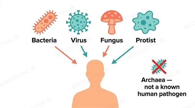
A flat diagram showing four pathogen categories in a row, each with a distinct icon, labeled Bacteria, Virus, Fungus and Protist, above a human silhouette, with a separate crossed-out Archaea icon labeled Archaea not a known human pathogen
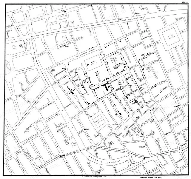
A real 1854 historical map by John Snow showing clustered cholera deaths in the Broad Street area of London around a single contaminated water pump
John Snow, 1854 · Public domain · Wikimedia Commons
C3.2.1 · NOS

Careful observation, before germ theory

Long before pathogens could be seen under a microscope, careful observation of disease patterns alone drove real progress against infection — a key nature-of-science idea in this topic.
  • In 1840s Vienna, Ignaz Semmelweis noticed childbed fever was far more common on wards where doctors came straight from autopsies, and cut deaths sharply just by requiring handwashing.
  • In 1854 London, John Snow traced a cluster of cholera deaths to one contaminated water pump on Broad Street by carefully mapping cases, establishing water-borne transmission through observation alone.
Why it mattersNeither breakthrough required identifying the causative microorganism — exam questions test what each scientist actually did, not a microbiology discovery neither of them made.
C3.2.2

Skin blocks two different ways

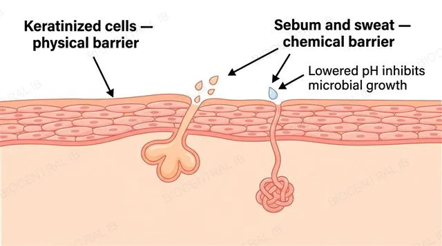
Skin acts as a primary defence against pathogens in two distinct ways at once: as a physical barrier and as a chemical barrier.
  • The physical barrier is the tightly packed, keratinised layer of dead cells at the skin's surface, which pathogens cannot easily penetrate.
  • The chemical barrier comes from secretions such as sebum and sweat, which lower the skin's surface pH and inhibit the growth of many microorganisms.
  • Both barriers act before any immune cell is even involved — pathogens are kept out, not fought off, at this first line of defence.
Why it mattersDescribing skin as only a physical barrier is a common error — its chemical, pH-based component is just as much a part of this same primary defence.
A flat cross-section diagram of skin showing a keratinized cell layer and a sebaceous gland secreting sebum onto the surface, labeled Keratinized cells physical barrier, Sebum and sweat chemical barrier, and Lowered pH inhibits microbial growth
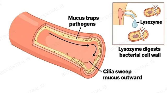
A flat diagram of an airway lined with mucous membrane, showing mucus trapping pathogen particles and cilia sweeping them outward, plus a small inset of a tear duct releasing lysozyme onto a bacterial cell wall, labeled Mucus traps pathogens, Cilia sweep mucus outward and Lysozyme digests bacterial cell wall
C3.2.2

Where skin can't seal the gap

Mucous membranes provide the same primary defence as skin, but at internal openings — the respiratory, digestive and reproductive tracts — that must stay open to function.
  • Because these tracts cannot be sealed like skin, mucous membranes trap incoming pathogens in sticky mucus, and structures such as cilia in the airway sweep trapped material back out.
  • Lysozyme, a chemical secretion present in tears and saliva, adds a further chemical barrier by digesting the cell walls of many bacteria.
Why it mattersMucous membranes are not simply weaker skin — they are a barrier suited to a structurally different job: sealing surfaces that must remain open.
C3.2.3

Sealing a cut, step by step

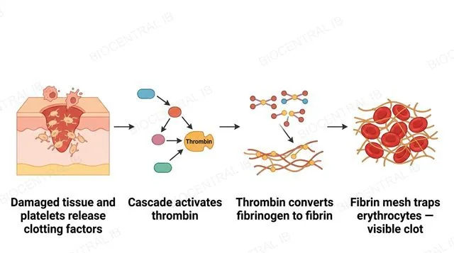
Blood clotting seals a cut in the skin through a cascade of reactions triggered by damaged tissue and activated platelets.
  • Damaged tissue and activated platelets at the wound release clotting factors, a cascade of proteins that activate each other in sequence.
  • This cascade converges on activating the enzyme thrombin, which catalyses the rapid conversion of the soluble plasma protein fibrinogen into insoluble fibrin.
  • The resulting fibrin mesh traps erythrocytes flowing through the wound site, and the fibrin plus trapped cells together form the visible clot.
Why it mattersNo further detail of the cascade — named clotting factors, or intrinsic/extrinsic pathways — is required; the overall sequence from damage to clot is what matters.
A flat four-stage diagram in left-to-right order: damaged tissue and platelets release clotting factors, clotting factor cascade activates thrombin, thrombin converts fibrinogen to fibrin, fibrin mesh traps erythrocytes forming a clot, with single-direction arrows and no other arrows
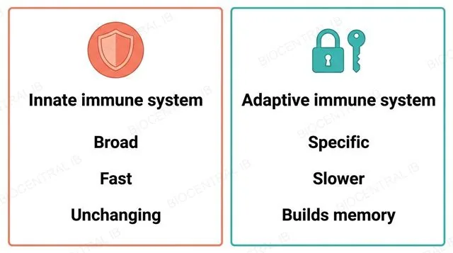
A flat two-panel comparison diagram, left panel labeled Innate immune system broad fast unchanging with a generic shield icon, right panel labeled Adaptive immune system specific slower builds memory with a lock-and-key icon, no other labels
C3.2.4

Two systems, two strategies

The innate and adaptive immune systems defend the body in fundamentally different ways, distinguished mainly by specificity and memory.
  • The innate system responds to broad categories of pathogen using the same mechanisms every time, and it does not change over an individual's lifetime.
  • The adaptive system responds specifically to a particular pathogen and builds up a memory of pathogens it has encountered, so its response becomes faster and more effective on repeated exposure.
  • This course requires no innate-system component beyond phagocytes — natural killer cells, interferons and the complement system are outside its scope.
Why it mattersMixing up which system is fast-but-broad versus slow-but-specific is the single most common error comparing these two systems.
C3.2.5

A cell that hunts by crawling

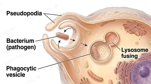
Phagocytes are innate immune cells that travel from the blood to a site of infection and destroy pathogens without any specificity for one particular pathogen type.
  • Phagocytes move from the bloodstream toward an infection site by amoeboid movement — flowing pseudopodia rather than swimming or crawling on legs.
  • On arrival, a phagocyte recognizes broad, shared features of a pathogen and engulfs it by endocytosis, enclosing it inside a vesicle.
  • Enzymes released from lysosomes then fuse with that vesicle and digest the pathogen — a purely innate mechanism with no antibody involvement.
Why it mattersThis lack of specificity and memory is exactly what places phagocyte defence in the innate, not adaptive, immune system.
A photorealistic labeled diagram of a phagocyte extending pseudopodia inward around a single bacterium and enclosing it into a phagocytic vesicle inside the cell, with a lysosome shown fusing onto that vesicle, labeled Pseudopodia, Bacterium pathogen, Phagocytic vesicle and Lysosome fusing, all arrows pointing inward toward engulfment with no outward or ambiguous arrows
Quick check

A person who has recovered from chickenpox rarely catches it again, but their phagocytes respond to every new infection in exactly the same broad way, no matter how many times they've encountered a pathogen before. What does this contrast illustrate?

The adaptive system builds memory and becomes more effective with exposure, while the innate system — including phagocytes — stays broad and unchanging
Phagocytes are actually part of the adaptive immune system
Memory is a feature of the innate immune system, not the adaptive system
Chickenpox immunity comes entirely from phagocyte activity
Correct answer: adaptive memory vs. unchanging innate response. Long-term immunity after an infection like chickenpox depends on adaptive-system memory cells; phagocytes are innate immune cells that respond the same broad way to every infection, with no memory or increase in effectiveness over time.
Part two

Adaptive immunity — the antibody response

C3.2.6 – C3.2.10
C3.2.6

One cell, one antibody

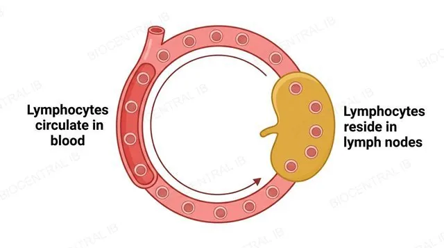
Lymphocytes are the cells of the adaptive immune system that cooperate to produce antibodies, circulating in the blood and also residing in lymph nodes.
  • Circulating through both the blood and lymph nodes means lymphocytes are well placed to encounter antigens draining from infected tissue almost anywhere in the body.
  • An individual has a very large number of distinct B-lymphocytes, and each one is committed to making just one specific type of antibody, matching one antigen shape.
  • This vast diversity of individually-committed B-cells means almost any antigen the body might meet has at least one matching B-cell already waiting to respond.
Why it mattersSpecificity here is per-cell, not general — no single B-cell can simply switch to making a different antibody on demand.
A flat diagram of a blood vessel connected to a lymph node, with lymphocyte icons shown moving along a circular path between the blood vessel and the lymph node, labeled Lymphocytes circulate in blood and Lymphocytes reside in lymph nodes
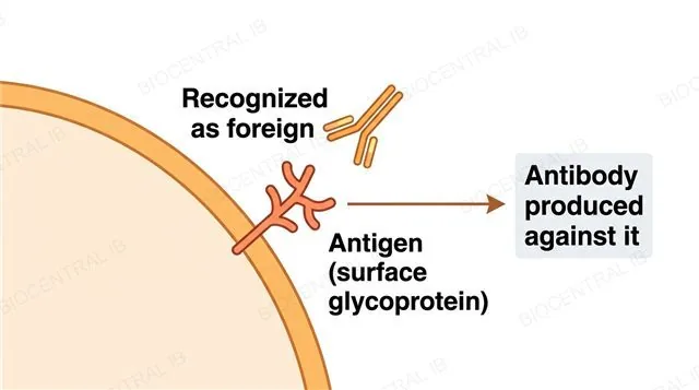
A flat diagram of a pathogen's outer surface with one glycoprotein antigen molecule labeled sticking outward, and a Y-shaped antibody approaching it from outside, labeled Antigen surface glycoprotein, Recognized as foreign and Antibody produced against it, a single arrow from antigen recognition to antibody production
C3.2.7

The tag that says foreign

An antigen is a recognition molecule that triggers antibody production because the adaptive immune system identifies it as foreign.
  • Most antigens are glycoproteins or other proteins, usually located on the outer surface of a pathogen, which is what makes them accessible for recognition.
  • Antigens are not unique to pathogens — surface antigens on erythrocytes can trigger antibody production if transfused into a person with a different blood group.
  • An antigen itself is not an antibody: the antigen is the foreign molecule being recognized, while the antibody is the protein later produced against it.
Why it mattersThe blood-transfusion example shows antigen recognition applies to any foreign surface molecule, not only ones arriving attached to a pathogen.
C3.2.8

Two signals, not one

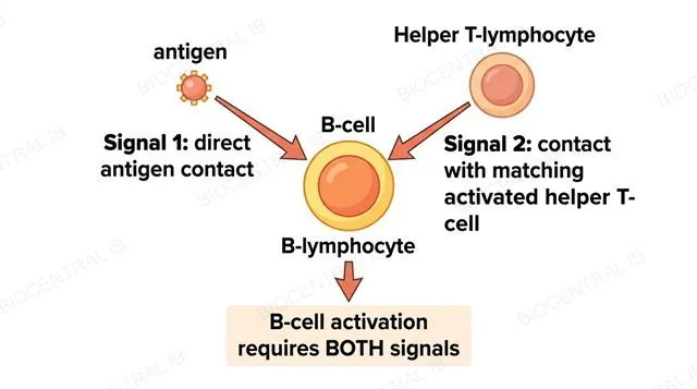
A B-lymphocyte only becomes fully activated — able to divide and produce antibodies — when it receives two separate signals together, not one alone.
  • The first signal is direct interaction between the B-cell and its one matching antigen; the second is contact with a helper T-lymphocyte that has itself been activated by that same antigen.
  • Both signals are required at once — antigen contact without a matching activated helper T-cell leaves the B-cell unactivated, and it will not proceed to divide.
  • This course does not require the detailed mechanism of helper T-cell activation itself, only that its activated contact is one of the two required signals.
Why it mattersMatching antigen specificity in both signals ensures the response actually matches the pathogen encountered.
A flat diagram of one B-lymphocyte receiving two separate arrows converging on it, one labeled Signal 1 direct antigen contact from an antigen molecule, one labeled Signal 2 contact with matching activated helper T-cell from a helper T-lymphocyte, both arrows meeting at a single box labeled B-cell activation requires both signals, no other arrows
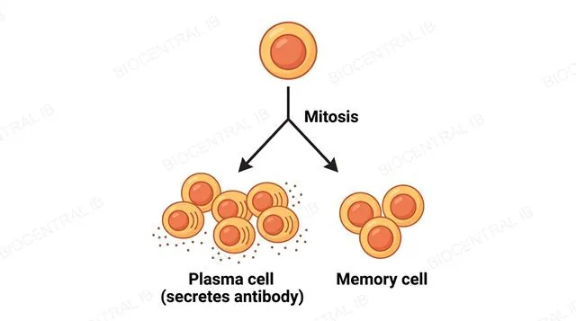
A flat diagram showing one activated B-lymphocyte at the top with a single downward arrow branching into a clone of several identical daughter cells below, most labeled Plasma cell secreting antibody and a smaller number labeled Memory cell, labeled Mitosis on the branching arrow
C3.2.9

From a few cells to an army

Once activated, a B-lymphocyte divides repeatedly by mitosis to form a clone of antibody-secreting plasma cells, all making the same antibody.
  • Only a relatively small number of B-cells match any one antigen, so this clonal division is what produces enough plasma cells to secrete a genuinely useful amount of antibody.
  • Division happens by mitosis, not any other process, producing genetically identical daughter cells rather than novel antibody variants.
  • Most of these daughter cells become plasma cells — short-lived antibody factories — while a smaller proportion become long-lived memory cells instead.
Why it mattersOne activated B-cell alone could never secrete enough antibody to matter; multiplying first is what makes the response effective.
C3.2.10

A response that remembers

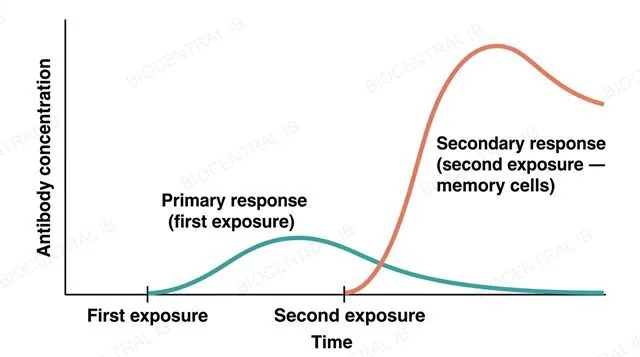
Immunity is the ability to eliminate an infectious disease from the body, and it exists because of the long-term survival of memory cells capable of making the specific antibody needed.
  • On first exposure, the primary response is relatively slow and reaches a low antibody concentration, often leaving time for symptoms to develop before the pathogen is cleared.
  • Memory cells persist for years afterward, so a second exposure triggers a secondary response that is faster, larger and reaches a higher antibody concentration.
  • This secondary response is usually fast enough to clear the pathogen before symptoms appear — the practical meaning of being immune.
Why it mattersVaccination deliberately triggers this same primary-response/memory-cell pathway, without the person ever experiencing the actual disease.
A flat line-graph diagram of antibody concentration against time after two exposures, showing a low slow-rising primary response curve after the first exposure labeled Primary response first exposure, and a distinctly higher, faster-rising secondary response curve after the second exposure labeled Secondary response second exposure memory cells, x-axis labeled Time and y-axis labeled Antibody concentration, the secondary curve clearly higher and faster than the primary curve with no other curves
Quick check

A B-cell binds its one matching antigen directly, but never encounters a matching activated helper T-cell. What happens?

The B-cell fails to fully activate and does not proceed to divide into plasma cells or memory cells
The B-cell still fully activates and starts secreting antibody
The B-cell becomes a phagocyte instead
The B-cell activates a completely unrelated B-cell clone
Correct answer: no activation without both signals. B-cell activation requires direct antigen interaction AND contact with a matching activated helper T-cell — antigen contact alone is not sufficient, so without the second signal the B-cell never proceeds to clonal division.
Part three

HIV, AIDS and antibiotics

C3.2.11 – C3.2.14
C3.2.11

One virus, specific routes

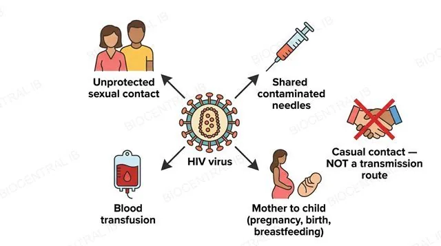
HIV (human immunodeficiency virus) is transmitted between people through the exchange of specific body fluids, not through casual everyday contact.
  • Recognised transmission routes include unprotected sexual contact, sharing contaminated needles or syringes, and transfusion of infected blood or blood products.
  • Mother-to-child transmission can also occur during pregnancy, during birth, or through breastfeeding — all involving exchange of body fluids between mother and child.
  • HIV is not transmitted by casual contact such as shaking hands, sharing food or drink, or airborne droplets — transmission always requires exchange of one of the specific fluids involved.
Why it mattersKnowing which routes are real and which are myths matters directly for public-health messaging, not just for the exam.
A flat diagram with a central HIV virus icon and four labeled arrows pointing outward to icons for Unprotected sexual contact, Shared contaminated needles, Blood transfusion and Mother to child pregnancy birth breastfeeding, plus one separate crossed-out icon for a handshake labeled Casual contact not a transmission route
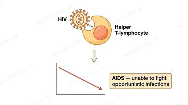
A flat diagram of an HIV virus icon attacking and destroying a helper T-lymphocyte specifically, with a downward arrow to a small line graph showing declining helper T-cell count over time, ending at a label AIDS unable to fight opportunistic infections, labeled HIV, Helper T-lymphocyte and AIDS unable to fight opportunistic infections, no other cell types shown as targets
C3.2.12

A virus that targets the helpers

HIV specifically infects and kills helper T-lymphocytes — not lymphocytes in general — and AIDS is the consequence of their numbers collapsing.
  • Because helper T-cell contact is one of the two required signals for B-cell activation, a falling helper T-cell count directly prevents new antibody responses from being mounted.
  • This leaves the body unable to fight infections it would normally control easily, so opportunistic infections that are rarely dangerous in a healthy person become life-threatening.
  • AIDS itself is not a separate infection — it is this state of collapsed adaptive immunity, caused by HIV's specific destruction of helper T-cells.
Why it mattersThe danger of AIDS comes from the opportunistic infections that follow immune collapse, not from HIV directly causing symptoms itself.
C3.2.13

Why antibiotics can't touch a virus

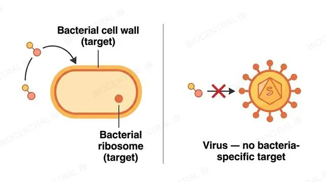
Antibiotics work by blocking specific processes that occur in bacterial cells but not in eukaryotic cells, giving them selective toxicity.
  • Targets include bacterial cell wall synthesis and the structurally distinct bacterial-type ribosome — processes human cells either lack entirely or carry out differently.
  • Because human cells don't rely on these same bacteria-specific structures, an antibiotic can disrupt bacteria without seriously harming the person taking it.
  • Viruses have no cell wall or ribosomes of their own — they hijack the host's eukaryotic machinery instead — leaving no bacteria-specific target for an antibiotic to block.
Why it matters"Antibiotics don't work on viruses because they're different" is too vague for full marks — the real reason is the total absence of a bacteria-specific target.
A flat two-panel diagram, left panel showing a bacterial cell with its cell wall and a distinct ribosome each labeled and an antibiotic molecule arrow blocking the cell wall, labeled Bacterial cell wall target and Bacterial ribosome target, right panel showing a virus with no cell wall and no ribosomes and one crossed-out antibiotic arrow, labeled Virus no bacteria-specific target
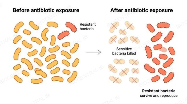
A flat two-panel before-and-after diagram in left-to-right order, left panel labeled Before antibiotic exposure showing a mixed population of bacteria icons mostly one color with a few differently colored resistant ones, right panel labeled After antibiotic exposure showing mostly the resistant colored bacteria remaining and reproducing, single left-to-right direction, labeled Sensitive bacteria killed and Resistant bacteria survive and reproduce
C3.2.14

Resistance evolves, it isn't learned

Resistance to antibiotics spreads through a bacterial population by natural selection acting on genetic variation that already exists, not by bacteria adapting during their own lifetime.
  • Antibiotic exposure is a selection pressure: pre-existing resistant variants survive and reproduce while susceptible bacteria are killed, so resistance becomes more common across generations.
  • Careful, judicious use of antibiotics — completing full courses, avoiding their use for viral infections — slows, though never fully prevents, the emergence of multi-resistant strains.
  • New techniques keep opening new research avenues: systematically screening large chemical libraries is one recent method yielding new candidate antibiotics.
Why it mattersDescribing bacteria as "learning" or "adapting" during exposure is the most common error here — resistance is selection on variation that was already there.
Part four

Public health — zoonoses, vaccines and herd immunity

C3.2.15 – C3.2.18
C3.2.15

When a disease crosses over

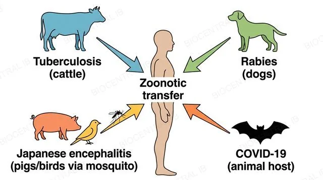
A zoonosis is an infectious disease that can transfer from another animal species into humans, and such diseases are a significant and varied part of human infectious disease.
  • Required examples include tuberculosis, rabies and Japanese encephalitis, each transferring from a different animal reservoir and by a different route of infection.
  • COVID-19 is included as a recent, large-scale example — SARS-CoV-2 is believed to have transferred from an animal host into humans, with profound consequences worldwide.
  • Not every disease with a non-human link is strictly zoonotic — malaria, for example, is vector-borne but not transferred from an animal reservoir the same way.
Why it mattersZoonoses illustrate why animal and human health are interdependent, not separate — a theme increasingly relevant to public-health policy.
A flat diagram with four separate animal-to-human transfer arrows, each labeled with one required example, Tuberculosis from cattle, Rabies from dogs, Japanese encephalitis from pigs and birds via mosquito, and COVID-19 from an animal host, each arrow pointing from its animal icon to a single human silhouette, labeled Zoonotic transfer
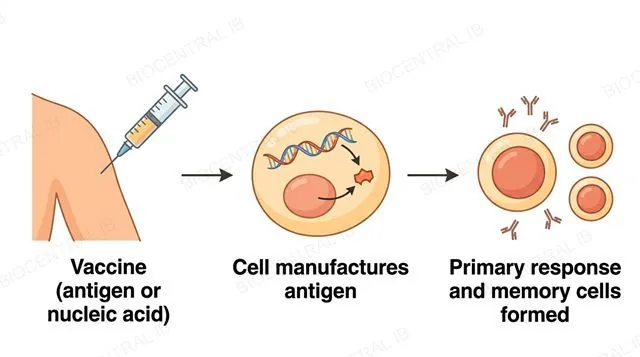
A flat left-to-right diagram, a syringe injecting an antigen or nucleic acid into an arm, an arrow to a body cell using the nucleic acid instructions to manufacture antigen, an arrow to an immune system icon producing antibodies and memory cells, labeled Vaccine antigen or nucleic acid, Cell manufactures antigen, Primary response and memory cells formed, single direction arrows only
C3.2.16

Training the immune system safely

A vaccine stimulates immunity to a specific pathogen without ever causing the disease itself, by introducing antigens, or the genetic instructions to make them.
  • A vaccine may contain the antigen directly, or nucleic acids (DNA or RNA) with sequences that code for that antigen, which the body's own cells then use to make it.
  • Either way, the immune system responds to the antigen as it would during a real infection — triggering a primary response and forming memory cells.
  • This achieves the same long-term protection as recovering from the disease, without the symptoms or transmission risk of actually being infected.
Why it mattersA vaccine supplies antigens, or instructions to make them — never ready-made antibodies — a frequent point of confusion worth fixing early.
C3.2.17

Protection that spreads beyond the individual

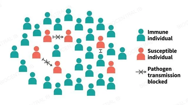
Herd immunity is the interdependence of members of a population in resisting a disease: if enough individuals are immune, transmission is greatly impeded even for those who aren't.
  • When a high enough percentage of a population is immune, a pathogen cannot easily find a continuous chain of susceptible hosts to sustain its spread.
  • This indirectly protects non-immune individuals — including those medically unable to be vaccinated — by lowering their real-world chance of exposure, not by making them personally immune.
  • Below this threshold, transmission chains reconnect and outbreaks can spread rapidly through the remaining susceptible population.
Why it mattersHerd immunity lowers exposure risk for the non-immune; it does not grant them personal immunity — a distinction worth stating explicitly.
A flat grid diagram of many small human silhouette icons, most colored to represent immune individuals and a few colored differently to represent susceptible individuals scattered among them, with short blocked arrows shown stopping between susceptible icons because immune icons separate them, labeled Immune individual, Susceptible individual and Pathogen transmission blocked
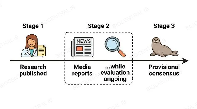
A flat three-stage left-to-right timeline diagram: a scientist publishing a study, a newspaper icon reporting on it while a separate ongoing-evaluation icon is shown still in progress at the same time, then a checkmark icon labeled provisional scientific consensus, labeled Research published, Media reports while evaluation ongoing and Provisional consensus, single left-to-right direction with the media-report and ongoing-evaluation icons clearly shown occurring at the same time not in sequence
C3.2.17 · NOS

Publishing is not the final word

Vaccine science carries specific nature-of-science lessons: scientists publish research precisely so other scientists can evaluate it, and that evaluation takes time.
  • Media often report on research while scientific evaluation is still ongoing, so consumers need to be aware a headline may precede the settled scientific picture.
  • Vaccines are tested unusually rigorously precisely because they are given to large numbers of healthy people — risks of side effects are minimal, but never nil.
  • Science generally works toward pragmatic, provisional truth rather than absolute certainty — a distinction often poorly understood outside science.
Why it mattersA published finding being provisional doesn't make it unreliable — it means it stays open to refinement as more evidence accumulates.
C3.2.18 · Skills

Two formulas, two different questions

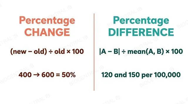
Evaluating COVID-19 pandemic data requires choosing the right calculation: percentage change for a genuine before/after pair, or percentage difference for two independent values.
  • Percentage change compares an old and new value of the same variable over time: (new − old) ÷ old × 100 — cases rising from 400 to 600 is a 50% change.
  • Percentage difference compares two independent values that aren't a before/after pair — such as two countries' same-day case rates — using |value 1 − value 2| ÷ mean × 100 instead.
  • Using percentage change's old-value denominator for two independent values instead is a common, specifically assessed error.
Why it mattersDeciding which formula applies is itself part of what this statement assesses, not just the arithmetic itself.
A flat two-column comparison infographic, left column headed Percentage change with the formula new minus old divided by old times 100 and the worked example 400 to 600 equals 50 percent, right column headed Percentage difference with the formula absolute value of A minus B divided by mean of A and B times 100 and the worked example 120 and 150 per 100000, no other formulas or numbers shown

Key vocabulary

Worth being able to define in a single sentence each

Pathogen
A disease-causing organism — typically a virus, bacterium, fungus or protist.
Antigen
A recognition molecule, usually a surface protein, that the adaptive immune system identifies as foreign.
Phagocyte
An innate immune cell that engulfs and digests pathogens by endocytosis, without specificity for one pathogen type.
Memory cell
A long-lived lymphocyte, formed after activation, responsible for a faster secondary immune response.
Herd immunity
Population-level protection that occurs when enough individuals are immune to impede a pathogen's transmission.
Zoonosis
An infectious disease that can transfer from another animal species into humans.
Vaccine
A preparation containing antigens, or instructions to make them, that stimulates immunity without causing disease.
Antibiotic resistance
The spread, by natural selection, of bacterial traits that survive antibiotic exposure.
AIDS
The immune collapse that follows HIV's destruction of helper T-lymphocytes.

Where this shows up again

B1.2
Antibodies are proteins with a variable region that determines their antigen specificity (B1.2). Explain how the structure of an antibody enables it to bind one specific antigen shape.
B2.1
Phagocytes engulf pathogens by endocytosis, a process that depends on the fluid, flexible nature of the plasma membrane (B2.1). Explain why the fluid mosaic structure of the membrane is necessary for endocytosis to occur.
A4.1
Antibiotic resistance in bacteria (C3.2.14) is an example of natural selection acting on a population (A4.1). Explain how the process of natural selection accounts for the spread of resistance through a bacterial population over time.

C3.2 Defence against disease — one-page recap

Screenshot this slide to revise from

Innate defence
  • Skin/mucous membranes (physical + chemical) and blood clotting act before any immune cell is involved.
  • Phagocytes engulf pathogens by endocytosis and digest them with lysosomal enzymes — broad, unchanging, no memory.
Innate vs adaptive
  • Innate: broad, fast, unchanging. Adaptive: specific, slower, builds memory and improves with exposure.
  • This course requires no innate component beyond phagocytes.
Antibody response
  • B-cell activation needs two signals — antigen contact + a matching activated helper T-cell.
  • Activated B-cells divide by mitosis into plasma cells (antibody factories) and memory cells (long-term immunity).
Primary vs secondary response
  • Primary: slow, low peak, first exposure. Secondary: fast, high peak, driven by memory cells.
  • Vaccines deliberately trigger this same pathway without causing the disease.
HIV, AIDS & antibiotics
  • HIV destroys helper T-cells specifically, collapsing antibody responses and causing AIDS.
  • Antibiotics block bacteria-specific processes — useless against viruses; resistance spreads by natural selection.
Public health
  • Zoonoses (tuberculosis, rabies, Japanese encephalitis, COVID-19) transfer from animals to humans.
  • Herd immunity lowers population-wide exposure; use percentage change (before/after) vs difference (independent values) correctly.

A body that remembers every enemy it has met

From a chemical barrier on skin to a memory cell waiting years for a second encounter, defence against disease is a system built to learn.
C3.2 Defence against disease · BioCentral IB
Use ↓ ↑ or click to navigate
01 / 33