BioCentral IBBioCentral IB
IB Biology · Theme D · D2.1

Cell and nuclear division

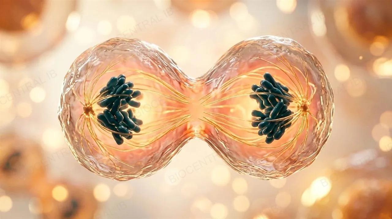
One cell becomes two. Mitosis copies a nucleus exactly, for growth and repair; meiosis halves the chromosome number and shuffles the genes, so the gametes differ genetically. This is how cells copy their DNA, share it out, and sometimes lose control of it.
Guiding questions

How can large numbers of genetically identical cells be produced?

How do eukaryotes produce genetically varied cells that can develop into gametes?

Part one

Dividing a cell

D2.1.1 – D2.1.4
D2.1.1

One mother cell, two daughter cells

In all living organisms, a parent cell, often called the mother cell, divides to produce two daughter cells.
  • Every cell comes from a cell that already existed, so every cell in your body traces back through a chain of divisions to one fertilized egg.
  • In a single-celled organism, division makes a whole new individual: a bacterium divides by binary fission into two bacteria.
  • In a multicellular organism, division supplies the extra cells needed for growth and replacements for damaged or worn-out cells.
  • In eukaryotes, division has two linked parts: nuclear division (mitosis or meiosis) and cytokinesis, the splitting of the cytoplasm.
Why it mattersThe rest of this topic asks two questions: how the DNA is shared out accurately, and how the cell then splits.
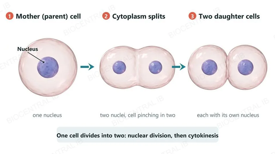
Three labeled stages of one animal cell dividing, left to right: a single round mother (parent) cell with one nucleus; the same cell pinched in the middle into two lobes, with two nuclei; and two round daughter cells, each with its own nucleus. Arrows lead from one stage to the next.
D2.1.2

Pinch or build: two ways to split the cytoplasm

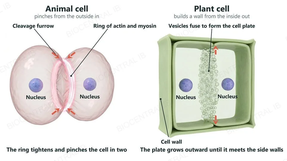
Cytokinesis is the splitting of the cytoplasm of a parent cell between its daughter cells.
  • Animal cell: a ring of the contractile proteins actin and myosin forms around the equator, just inside the plasma membrane. It tightens like a drawstring, pinching the cell in two.
  • Plant cell: the cell wall is too rigid to pinch. Vesicles carrying membrane and cell wall material gather at the equator and fuse into a new partition, the cell plate, which grows outward until it reaches the side walls.
  • Both methods end the same way: two daughter cells, each with its own nucleus and its own plasma membrane.
Why it mattersAnimal cells pinch from the outside in; plant cells build from the inside out.
Two labeled cells in cytokinesis. On the left, an animal cell pinched into two lobes by a glowing ring of actin and myosin around its waist, with a cleavage furrow at the surface, red arrows showing the groove moving inward, and one nucleus in each lobe. On the right, a plant cell inside a rigid wall, cut open, with a partition of fusing vesicles, the cell plate, growing outward from its middle toward the side walls, and a nucleus on each side.
D2.1.3

Splitting the cytoplasm unevenly

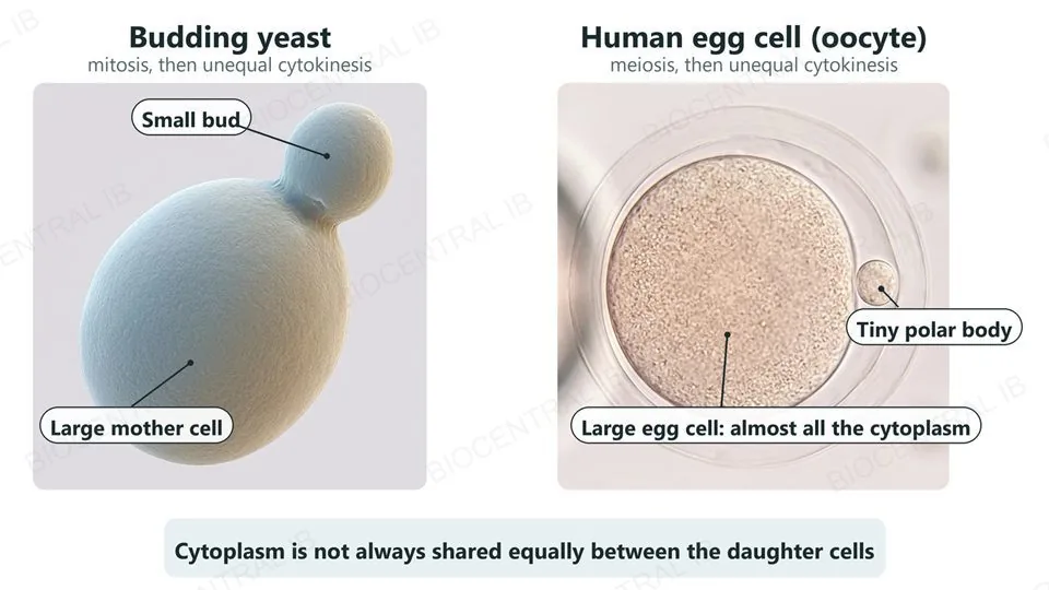
Cytokinesis usually shares the cytoplasm about equally between the daughter cells, but in some cases it does not.
  • Either way, both daughter cells must receive at least one mitochondrion, and any other organelle that can only be made by dividing a pre-existing one, because a cell cannot build one from scratch.
  • Budding in yeast: a small bud grows out of the mother cell, receives one of the two nuclei after mitosis, and pinches off as a much smaller daughter cell.
  • Oogenesis in humans: meiosis splits the cytoplasm very unevenly, so the large egg cell keeps almost all of it and a tiny polar body gets a nucleus but almost no cytoplasm.
Why it mattersThe egg keeps the stores of nutrients and organelles that the early embryo will need.
Two labeled examples of unequal cytokinesis: a scanning-electron-micrograph-style budding yeast, with a large mother cell and a much smaller bud; and a micrograph-style human egg cell (oocyte) that keeps almost all the cytoplasm, with a tiny polar body beside it inside its clear shell.
D2.1.4

Two kinds of nuclear division

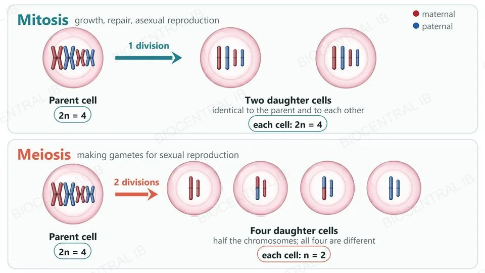
In eukaryotes, mitosis and meiosis are the two types of nuclear division, and each has a different job.
  • Nuclear division must happen before the cytoplasm splits. Otherwise one daughter cell could receive no nucleus at all: an anucleate cell, with no DNA to direct it.
  • Mitosis maintains the chromosome number and the genome: both daughter nuclei have the same chromosomes and genes as the parent. It is used for growth, tissue repair and asexual reproduction.
  • Meiosis halves the chromosome number and generates genetic diversity. It is used to make gametes for sexual reproduction.
Why it mattersMitosis makes copies; meiosis halves and shuffles. Everything that follows builds on this contrast.
Two rows of cells. Top row, mitosis: a parent cell with four chromosomes (two homologous pairs, maternal red and paternal blue) divides once into two daughter cells that each have the same four chromosomes as the parent and each other. Bottom row, meiosis: a parent cell with the same four chromosomes divides twice into four daughter cells that each have only two chromosomes, one of each pair, and all four cells differ from each other because some chromosomes carry swapped red and blue segments.
Part two

Chromosomes: copied, coiled and moved

D2.1.5 – D2.1.6
D2.1.5

Copy the DNA first

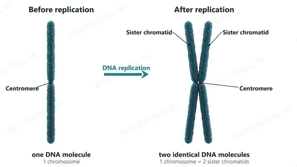
DNA replication is a prerequisite for both mitosis and meiosis: it comes first, so that there is enough DNA for each daughter nucleus to receive a full set.
  • After replication, each chromosome consists of two identical DNA molecules, called sister chromatids, made by copying the same original molecule.
  • The sister chromatids stay held together, especially at the centromere, until anaphase, when they separate.
  • Once separated, each chromatid is a chromosome in its own right and travels to one of the two new nuclei.
Why it mattersSkip replication and each daughter nucleus would receive only half of the parent's DNA.
Two textured chromosomes side by side, labeled. On the left, before replication, a single thin rod made of one DNA molecule with a narrow constriction, the centromere. On the right, after replication, one chromosome made of two identical sister chromatids joined at the centromere, forming a slim X.
D2.1.6

Coil it up: condensation

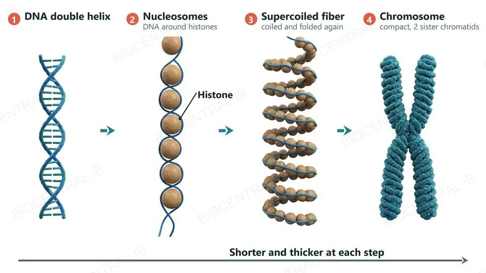
Before division, each long, thin DNA molecule is condensed into a short, compact chromosome that can be moved without tangling or breaking.
  • Histones: DNA is wound around histone proteins, forming nucleosomes that look like beads on a string.
  • Supercoiling: this beaded fiber is coiled and folded again and again, with the help of proteins such as condensin, into a chromosome many times shorter and thicker.
  • Condensed chromosomes can be seen with a light microscope. In interphase the DNA is spread out as chromatin and separate chromosomes cannot be seen.
Why it mattersLong, thin, uncondensed DNA would tangle and snap when it was pulled apart.
Four numbered stages of DNA condensation from left to right: a DNA double helix; DNA wound around histone protein beads (nucleosomes) like beads on a string; the beaded fiber coiled and folded into a thick spiral, the supercoiled fiber; and a short, thick, compact chromosome made of two sister chromatids. A long arrow underneath reads shorter and thicker at each step.
D2.1.6

Pulled apart by microtubules

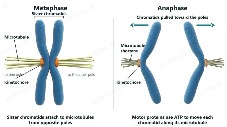
Chromosomes are moved by microtubules, which make up the spindle, and by microtubule motor proteins.
  • Spindle microtubules grow from the two poles of the cell and attach to a protein disc, the kinetochore, at the centromere of each sister chromatid.
  • In mitosis, the two sister chromatids attach to microtubules from opposite poles, so that when they separate they are pulled in opposite directions.
  • Motor proteins use energy from ATP to move chromosomes along the microtubules, and the microtubules shorten at the kinetochore as each chromatid is drawn toward its pole.
Why it mattersChromosome movement is one of the life processes that ATP powers (C1.2).
Two labeled views of one chromosome and its spindle microtubules. Left, metaphase: an X-shaped chromosome whose two sister chromatids each carry an orange kinetochore facing opposite directions, each attached to a bundle of microtubules running to its own spindle pole. Right, anaphase: the chromatids have separated; each is bent like a V with its kinetochore at the tip pointing toward its own pole, attached to a shorter bundle of microtubules, and is being pulled toward that pole.
Part three

Mitosis

D2.1.7 – D2.1.8
D2.1.7

Four phases, two identical nuclei

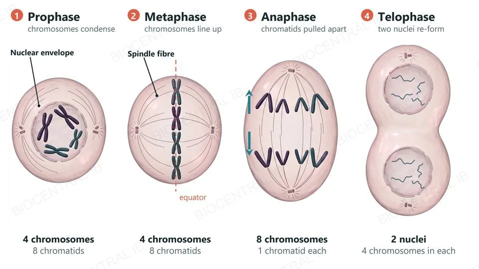
Mitosis is a continuous process, divided for study into four phases: prophase, metaphase, anaphase and telophase.
  • Prophase: chromosomes condense and become visible, each as two sister chromatids; the spindle begins to form and the nuclear envelope breaks down.
  • Metaphase: the chromosomes line up at the equator, each attached to spindle microtubules from both poles.
  • Anaphase: the sister chromatids separate and are pulled to opposite poles; each is now a chromosome.
  • Telophase: chromosomes reach the poles and uncoil, nuclear envelopes re-form and the spindle disappears. Cytokinesis, which usually begins during telophase, then splits the cell.
Why it mattersIdentical chromatids go to each pole, so both daughter nuclei are genetically identical to the parent.
Four cells in a row showing the phases of mitosis in a cell with four chromosomes, numbered 1 to 4. Prophase: four condensed X-shaped chromosomes, eight chromatids, inside a breaking-down nuclear envelope. Metaphase: the four chromosomes lined up along the equator on the spindle. Anaphase: eight single chromatids, four moving to each pole, now counted as eight chromosomes. Telophase: an elongated cell pinching in two, with two new nuclei that each hold four chromosomes.
D2.1.7

Count chromosomes and chromatids

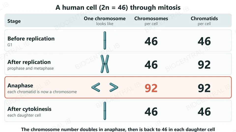
Counting through mitosis is a common exam trap, because a chromosome has two chromatids only from replication until anaphase. Take a human cell, with 46 chromosomes.
  • Before replication: 46 chromosomes, each one DNA molecule.
  • After replication, through prophase and metaphase: still 46 chromosomes, but each has two sister chromatids, so there are 92 chromatids.
  • Anaphase: each separated chromatid is now counted as a chromosome, so the cell briefly has 92 chromosomes, 46 moving to each pole.
  • After telophase and cytokinesis: each daughter cell has 46 chromosomes, the same number and the same genes as the parent.
Why it mattersThe chromosome number doubles in anaphase and is back to 46 once the cell divides; the DNA content doubled earlier, at replication.
A table of chromosome and chromatid counts for a human cell through mitosis, with a picture of a chromosome at each stage. Before replication: 46 chromosomes and 46 chromatids. After replication, in prophase and metaphase: 46 chromosomes and 92 chromatids. In anaphase, highlighted: 92 chromosomes and 92 chromatids, each separated chromatid now counted as a chromosome. In each daughter cell after cytokinesis: 46 chromosomes and 46 chromatids.
D2.1.8

Which phase is this?

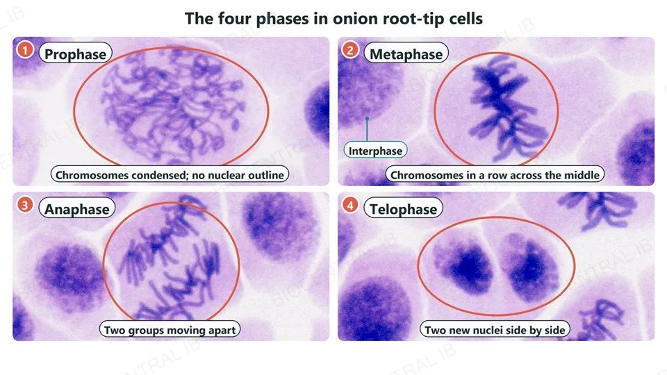
To identify a phase of mitosis in a diagram or micrograph, look at the chromosomes, the nuclear envelope and the shape of the cell.
  • Prophase: condensed chromosomes visible as threads or X shapes scattered through the cell, with no clear nuclear outline.
  • Metaphase: chromosomes lined up in a single row across the middle of the cell.
  • Anaphase: two groups of V-shaped chromatids moving apart, and the cell is lengthening.
  • Telophase: two separate clusters of chromosomes at opposite ends, which become two new nuclei, often with a cell plate or a pinching furrow between them.
Why it mattersIn a root-tip squash, most cells show an intact nucleus and no visible chromosomes (interphase); only a few are caught in mitosis.
Four real light-microscope photographs of stained onion root-tip cells, numbered 1 to 4, each with the dividing cell ringed. Prophase: condensed thread-like chromosomes and no nuclear outline. Metaphase: the chromosomes in a row across the middle of the cell, with a nearby interphase nucleus labeled. Anaphase: two groups of chromatids moving apart. Telophase: two new nuclei side by side.
J. Reischig · CC BY-SA 3.0 · Wikimedia Commons
Part four

Meiosis and genetic variation

D2.1.9 – D2.1.11
D2.1.9

One diploid nucleus, four haploid nuclei

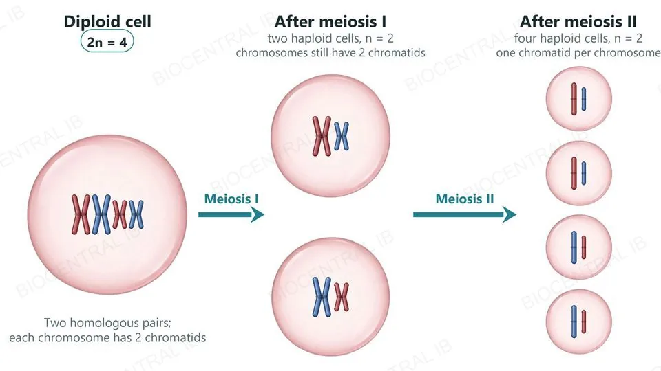
Meiosis is a reduction division: two divisions in a row turn one diploid nucleus into four haploid nuclei.
  • A diploid (2n) nucleus has its chromosomes in homologous pairs, one of each pair from each parent; a haploid (n) nucleus has just one chromosome of each pair. In humans, 2n = 46 and n = 23.
  • DNA is replicated once, before meiosis I. Meiosis I and meiosis II then follow, each with its own prophase, metaphase, anaphase and telophase (for example, metaphase I).
  • Meiosis I halves the chromosome number, giving two haploid nuclei; meiosis II divides each of those again, giving four haploid nuclei in all.
Why it mattersDNA is copied once but divided twice, so the chromosome number is halved.
Three columns showing meiosis in a cell with four chromosomes, two homologous pairs (red maternal, blue paternal). One diploid cell (2n = 4) whose chromosomes each have two chromatids; two haploid cells after meiosis I (n = 2), each with two chromosomes that still have two chromatids; and four haploid cells after meiosis II (n = 2), each with two single-chromatid chromosomes. Arrows are labeled meiosis I and meiosis II.
D2.1.9

Two rounds of segregation

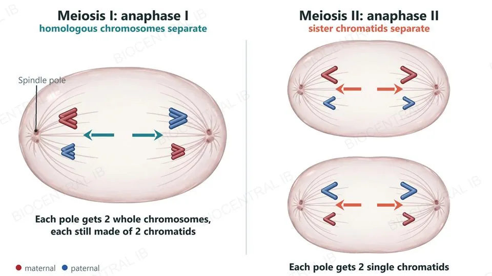
Meiosis I separates whole homologous chromosomes; meiosis II separates sister chromatids.
  • In prophase I, homologous chromosomes pair up as bivalents. In metaphase I, the bivalents line up at the equator. In anaphase I, the two chromosomes of each pair are pulled to opposite poles.
  • Each chromosome still has two sister chromatids after meiosis I, and there is no further DNA replication before meiosis II.
  • Meiosis II resembles mitosis: the chromosomes line up at the equator in metaphase II, then the sister chromatids separate in anaphase II and move to opposite poles.
Why it mattersMeiosis I is the reduction division: it is where diploid (2n) becomes haploid (n).
Two labeled views of the separations in meiosis. Left, anaphase I: in one cell, whole chromosomes that each still have two chromatids are pulled to opposite poles, two to each pole, so the homologous chromosomes separate. Right, anaphase II: in each of two cells, single chromatids are pulled to opposite poles, two to each pole, so the sister chromatids separate.
D2.1.9

Why sex needs meiosis

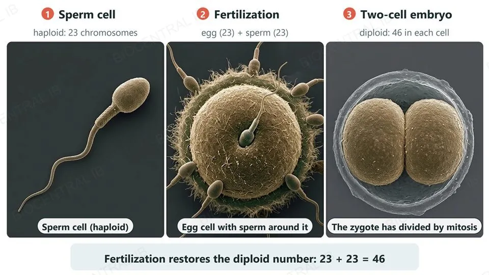
In a sexual life cycle, two gametes fuse at fertilization, so meiosis is needed to halve the chromosome number of each gamete.
  • Human body cells are diploid, with 46 chromosomes. Meiosis makes gametes, sperm and egg cells, that are haploid, with 23 chromosomes each.
  • At fertilization the sperm and egg nuclei fuse, and the zygote has 23 + 23 = 46 chromosomes: the diploid number is restored.
  • Without meiosis, the chromosome number would double in every generation.
  • Meiosis also breaks up the parents' combinations of alleles, so fertilization can produce new combinations.
Why it mattersThe zygote then divides by mitosis, so every body cell inherits the same 46 chromosomes.
Three numbered scanning-electron-micrograph-style views: a single human sperm cell, haploid with 23 chromosomes; a human egg cell with sperm around it at fertilization; and a two-cell early embryo, diploid with 46 chromosomes in each cell. Fertilization restores the diploid number: 23 plus 23 makes 46.
D2.1.10

When chromosomes fail to separate

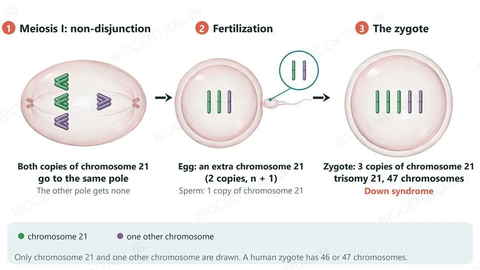
Non-disjunction is the failure of chromosomes or chromatids to separate in meiosis, so gametes receive too many or too few chromosomes.
  • If both chromosomes of a pair go to the same pole in anaphase I (or both chromatids in anaphase II), one gamete gets an extra chromosome and another gets none.
  • If a gamete with two copies of chromosome 21 fuses with a normal gamete, the zygote has three (trisomy 21) and 47 chromosomes: Down syndrome.
  • Most trisomy 21 arises in the mother's meiosis, and the risk rises with maternal age: about 1 in 1,500 births at age 20 and 1 in 350 at age 35.
Why it mattersA zygote with only one copy of chromosome 21 rarely survives, so it is trisomy that is seen in live births.
Three numbered stages. 1, meiosis I: an elongated cell in anaphase I in which both copies of chromosome 21 (green) move to the same pole and the other pole gets none. 2, fertilization: an egg with two copies of chromosome 21 (n + 1) meets a sperm with one copy. 3, the zygote has three copies of chromosome 21, trisomy 21, and 47 chromosomes: Down syndrome. A key shows chromosome 21 in green and one other chromosome in purple; only these two are drawn.
D2.1.10

Seeing trisomy 21 in a karyotype

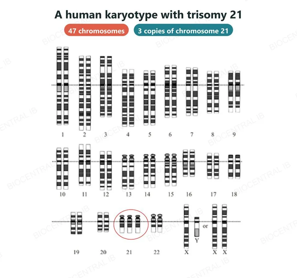
A karyotype is an image of a person's chromosomes, arranged in homologous pairs by size and banding pattern, so an extra or missing chromosome shows up at a glance.
  • The 22 pairs of autosomes are numbered roughly from largest (1) to smallest (22); the last pair are the sex chromosomes, shown here as X and Y (male) or two X (female).
  • Here chromosome 21 appears three times instead of twice, so the cell has 47 chromosomes: trisomy 21, the cause of Down syndrome.
  • Karyotypes of fetal cells, collected by amniocentesis or chorionic villus sampling, can reveal non-disjunction before birth.
Why it mattersA karyotype turns an invisible meiosis error into something a clinician can count.
A human karyotype from a person with Down syndrome: the 22 pairs of autosomes, numbered 1 to 22 and drawn with their banding patterns, then the sex chromosomes (X and Y, or two X). Chromosome 21 appears three times instead of twice, ringed in red, giving 47 chromosomes in all. Two labels read 47 chromosomes and 3 copies of chromosome 21.
Karyotype: NHGRI (adapted) · CC BY-SA 4.0 · Wikimedia Commons
D2.1.11

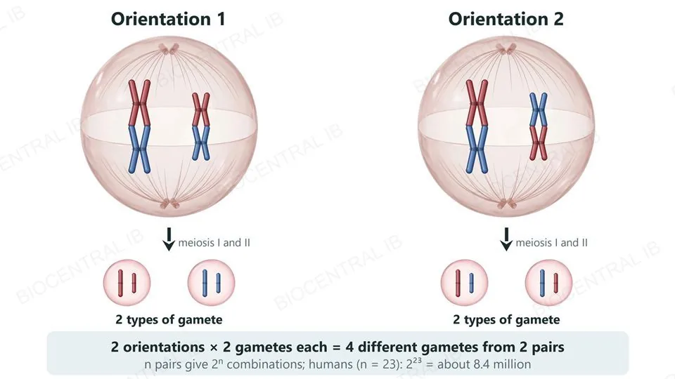
Random orientation shuffles the parents

At metaphase I, each bivalent lines up at the equator, and which chromosome faces which pole is random and independent of every other bivalent.
  • A bivalent's maternal and paternal chromosomes can face either pole, so each pole can receive any mixture of maternal and paternal chromosomes.
  • With n pairs of chromosomes there are 2n possible combinations. Humans, with n = 23, can make 223, about 8.4 million, different gametes from random orientation alone.
  • Two bivalents can line up in two ways and give four different types of gamete (each way gives two).
  • Random orientation is the physical basis of independent assortment of genes on different chromosomes (D3.2).
Why it mattersFertilization then joins two such gametes at random, multiplying the variety further.
Two identical cells at metaphase I, each with two bivalents (a long pair and a short pair of homologous chromosomes, red maternal and blue paternal) lined up at the equator between two spindle poles. In orientation 1 both red chromosomes face the top pole; in orientation 2 the long red chromosome faces the top pole and the short red one faces the bottom pole. Below each cell are the two types of gamete it produces, four different gamete types in all. A note gives 2 to the power n combinations, about 8.4 million in humans.
D2.1.11

Crossing over: swapping segments

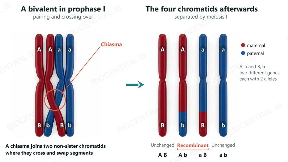
Crossing over is the exchange of matching segments of DNA between non-sister chromatids of homologous chromosomes during prophase I.
  • Homologous chromosomes pair up as a bivalent, and non-sister chromatids become linked at points called chiasmata.
  • At a chiasma, two chromatids break at matching points and rejoin crosswise, swapping the segments beyond it.
  • The two chromatids involved now carry a mixture of maternal and paternal alleles: new combinations that were not together in either parent's chromosome.
  • The sister chromatids are no longer identical, so the chromatids that separate in meiosis II differ from each other too.
Why it mattersCrossing over mixes alleles within a chromosome; random orientation shuffles whole chromosomes. Together they make each gamete unique.
Two views of a pair of homologous chromosomes, one red (maternal) and one blue (paternal), each made of two sister chromatids, with two genes marked A/a and B/b. Left: the pair lies side by side and two non-sister chromatids cross at one chiasma near the bottom. Right: the four chromatids after the exchange: red A B unchanged, red A with a blue lower end b (recombinant), blue a with a red lower end B (recombinant), and blue a b unchanged.
Quick check

A human cell (2n = 46) has just finished meiosis I. How many chromosomes does each of the two new cells contain, and how many chromatids does each chromosome have?

46 chromosomes, each made of two sister chromatids
23 chromosomes, each made of two sister chromatids
23 chromosomes, each made of one chromatid
46 chromosomes, each made of one chromatid
Correct answer: 23 chromosomes, each with two chromatids. Meiosis I separates the two chromosomes of each homologous pair, so each cell gets 23 (one from every pair). It does not separate sister chromatids, so each chromosome still has two chromatids until anaphase II.

Key vocabulary — division, mitosis and meiosis

Worth being able to define in a single sentence each

Cytokinesis
The splitting of the cytoplasm of a parent cell between its daughter cells.
Sister chromatids
The two identical DNA molecules of a replicated chromosome, held together until anaphase.
Histone
A protein that DNA winds around; histones and supercoiling condense DNA into chromosomes.
Mitosis
Nuclear division that gives two genetically identical nuclei with the same chromosome number as the parent.
Diploid (2n) and haploid (n)
Diploid: chromosomes in homologous pairs. Haploid: one chromosome of each pair.
Bivalent
A pair of homologous chromosomes lined up together in meiosis I.
Crossing over
The exchange of segments between non-sister chromatids of homologous chromosomes in prophase I.
Non-disjunction
The failure of chromosomes or chromatids to separate in meiosis, giving gametes with too many or too few chromosomes.
Meiosis
A reduction division: two nuclear divisions that turn one diploid nucleus into four genetically varied haploid nuclei.
Part five · HL

HL — the cell cycle, its control and cancer

D2.1.12 – D2.1.17
D2.1.12 · HL

Proliferation for growth, replacement and repair

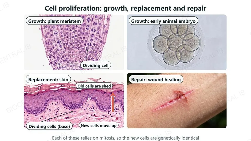
Cell proliferation, the production of many new cells by repeated cell division, supports growth, cell replacement and tissue repair.
  • Growth: plant meristems, at the tips of roots and shoots, keep dividing so the plant lengthens. In an early animal embryo, rapid divisions turn one fertilized egg into a ball of many cells.
  • Replacement: cells at the base of the skin's outer layer keep dividing, and their daughter cells move up to replace the surface cells that are constantly shed, renewing the outer layer about every month.
  • Repair: after a cut, cells at the edge of the wound divide and fill the gap.
Why it mattersAll of these rely on mitosis, which makes genetically identical cells.
Four labeled photographs of cell proliferation: a plant root tip where meristem cells are dividing, for growth; an early animal embryo of eight cells, for growth; a section of skin with dividing cells at the base, new cells moving up and old surface cells being shed, for replacement; and a healing cut on an arm, for repair.
D2.1.13 · HL

The cell cycle: grow, copy, divide

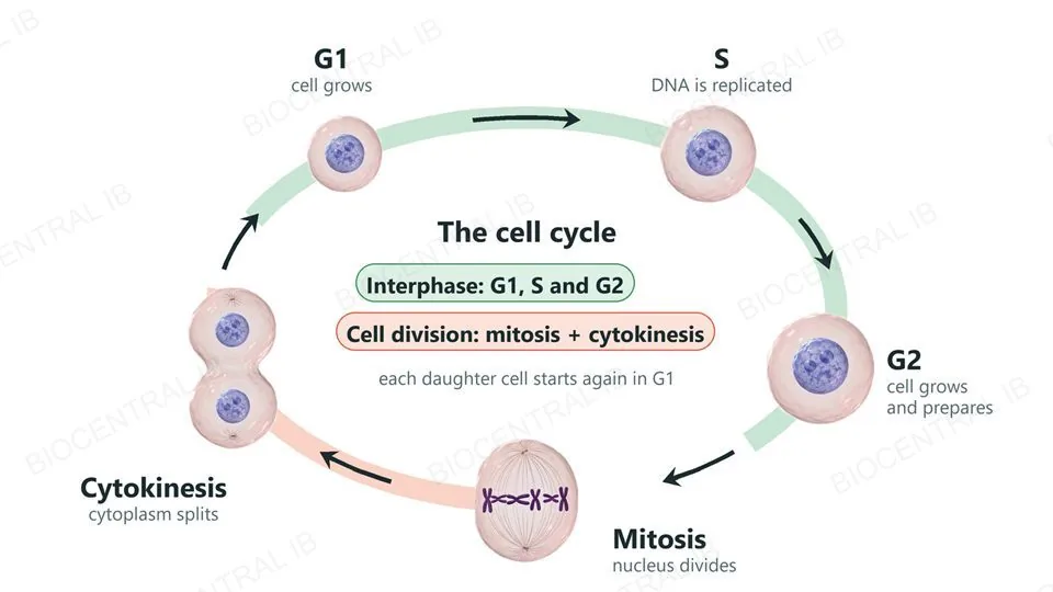
The cell cycle is the ordered sequence of events by which cells proliferate: interphase (G1, S and G2), then mitosis, then cytokinesis.
  • G1 (first gap): the cell grows and carries out its normal functions.
  • S (synthesis): the cell replicates its DNA, so every chromosome now has two sister chromatids.
  • G2 (second gap): the cell keeps growing and prepares for mitosis.
  • Mitosis, then cytokinesis, complete the cycle, and each daughter cell begins in G1.
Why it mattersIn a human cell that divides about once a day, mitosis takes only around an hour: most of the cycle is interphase.
A ring showing the cell cycle. G1: a small cell with a nucleus, the cell grows. S: a medium cell, DNA is replicated. G2: a larger cell that grows and prepares. Mitosis: a cell with condensed chromosomes on a spindle. Cytokinesis: a cell pinching into two, with arrows leading back to G1. G1, S and G2 make up interphase; mitosis and cytokinesis make up cell division.
D2.1.14 · HL

Interphase is not resting

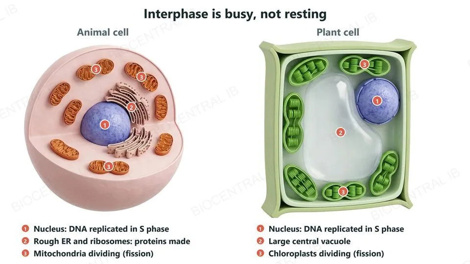
Interphase is a metabolically active period, during which the cell grows by making new cell components.
  • Growth involves biosynthesis: the cell makes proteins (by transcription and translation) and other components, and replicates its DNA in S phase.
  • Mitochondria, and chloroplasts in plant cells, increase in number by growing and dividing, because they can only arise from existing organelles.
  • Growth in G1 and G2 means the daughter cells are not smaller and smaller at each division.
Why it mattersInterphase is a common trap: it is not a resting phase, it is when most of the cell's work for the next division is done.
Two cut-open interphase cells with numbered labels. Left, an animal cell: 1 the nucleus, where DNA is replicated in S phase; 2 rough endoplasmic reticulum with ribosomes, where proteins are made; and 3 the three mitochondria that are pinching into two among the others. Right, a plant cell: 1 the nucleus; 2 a large central vacuole; and 3 chloroplasts, two of which are pinching into two.
D2.1.15 · HL

Cyclins decide whether the cycle goes on

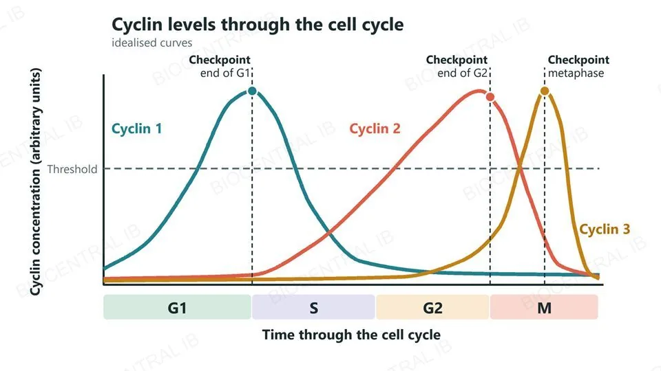
Cyclins are proteins whose concentrations rise and fall in a regular pattern through the cell cycle and control progress through it.
  • The cycle has checkpoints, such as at the end of G1, the end of G2 and in metaphase, where the cell continues only if conditions are satisfactory.
  • To pass a checkpoint, a specific cyclin must reach a threshold concentration. Different checkpoints need different cyclins, and each falls again as the cycle moves on.
  • Cyclins work by activating enzymes called cyclin-dependent kinases, which trigger the next stage. The roles of individual cyclins are not needed.
Why it mattersIf the threshold is not reached, the cell does not pass the checkpoint, so damaged or unready cells are held back.
A graph of cyclin concentration against time through G1, S, G2 and M, with three illustrative curves, cyclin 1, cyclin 2 and cyclin 3, that each rise to a peak and fall at different stages; a dashed horizontal threshold line; and three checkpoints, at the end of G1, the end of G2 and in metaphase, where the relevant cyclin is above the threshold.
D2.1.16 · HL

When the accelerator sticks or the brake fails

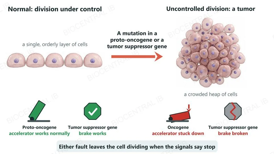
Mutations in genes that control the cell cycle can cause uncontrolled cell division, the basis of tumor formation.
  • A proto-oncogene is a normal gene that stimulates cell division. A mutation can convert it into an oncogene, which is overactive and drives division even when it should not: an accelerator stuck down.
  • A tumor suppressor gene normally restrains cell division. A mutation that disables it removes the brake.
  • Either kind of mutation can lead to uncontrolled division and a tumor; cancers usually follow several such mutations in one cell line.
  • Examples: RAS is a proto-oncogene, mutated in about one in five human cancers; p53 is a tumor suppressor gene, mutated in about half.
Why it mattersBoth routes leave the cell dividing when the normal signals say stop.
Left, a single orderly layer of five cells under normal control, with a working accelerator (a proto-oncogene) and a working brake (a tumor suppressor gene). Right, after a mutation, a crowded heap of many cells, with an accelerator stuck down (an oncogene) and a broken brake (a disabled tumor suppressor gene). Either fault leaves the cell dividing when the signals say stop.
D2.1.17 · HL

Benign or malignant: what a tumor does

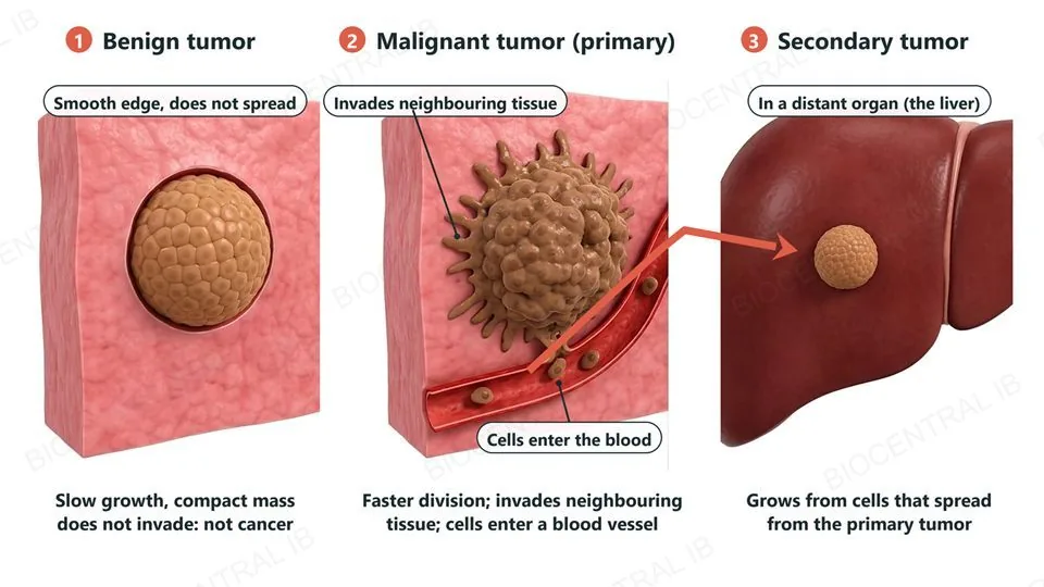
A tumor is an abnormal mass of cells produced by uncontrolled division; tumors differ in how fast they grow and whether they invade.
  • A benign tumor grows slowly, stays as a compact mass in one place and does not invade neighbouring tissue or spread. It is not cancer, though it can harm by pressing on organs.
  • A malignant tumor divides faster, invades neighbouring tissue and can metastasize: cells break away, travel in the blood or lymph, and start new tumors elsewhere. Malignant tumors are cancer.
  • The original tumor is the primary tumor; one that grows from cells that spread from it is a secondary tumor.
Why it mattersMost cancer deaths are caused by secondary tumors, not the primary one.
Three numbered cut-away views: a benign tumor, a compact rounded mass with a smooth edge that does not invade; a malignant primary tumor with an irregular edge invading the neighbouring tissue, with cells entering a blood vessel; and a small secondary tumor in a distant organ, the liver, with an arrow from the primary tumor along the blood vessel to it.
D2.1.17 · HL

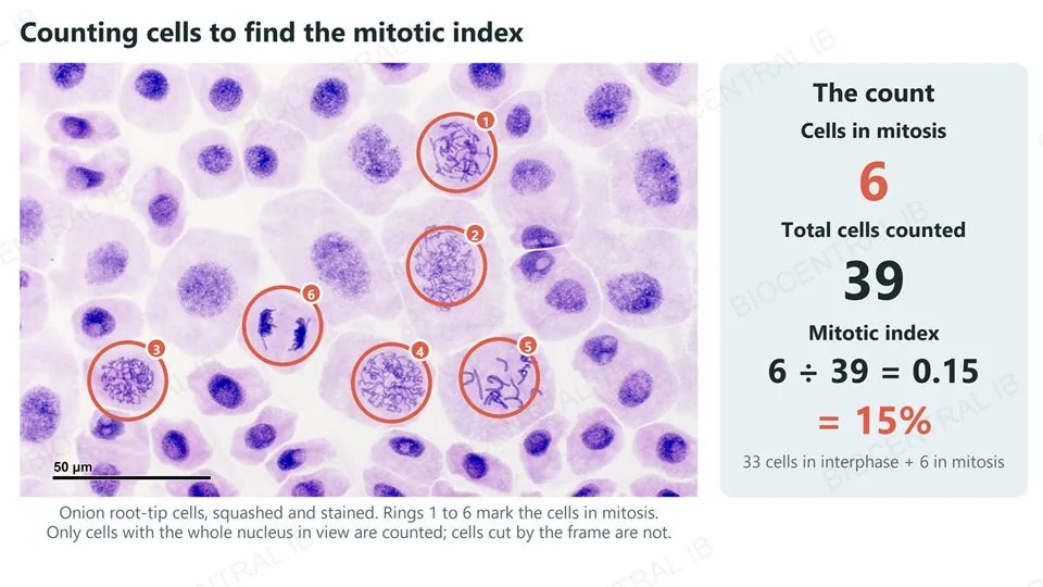
The mitotic index: counting dividing cells

The mitotic index is the proportion of cells in a tissue sample that are undergoing mitosis.
  • Mitotic index = number of cells in mitosis ÷ total number of cells counted, given as a decimal or a percentage.
  • Count every cell in the field of view: cells in any phase of mitosis are dividing; the rest are in interphase. Example: 12 dividing cells among 150 cells gives 12 ÷ 150 = 0.08, or 8%.
  • A higher index means a higher rate of cell division. Tumor tissue often has a higher mitotic index than normal tissue of the same type.
  • Count several fields, because one field is a small sample.
Why it mattersThe index is a snapshot: each cell spends only a small part of its cycle in mitosis.
A real micrograph of stained onion root-tip cells with the six cells in mitosis ringed and numbered 1 to 6, and a count box: 6 cells in mitosis among 39 cells counted gives a mitotic index of 6 divided by 39, which is 0.15 or 15 percent. Only cells with the whole nucleus in view were counted.
J. Reischig · CC BY-SA 3.0 · Wikimedia Commons
Quick check · HL

Which pair of statements correctly describes how mutations in cell-cycle control genes cause uncontrolled cell division?

Both a proto-oncogene and a tumor suppressor gene must become overactive
A proto-oncogene mutates into an overactive oncogene, and a tumor suppressor gene mutates so that it stops working
A proto-oncogene mutates so that it stops working, and a tumor suppressor gene mutates into an overactive oncogene
Both a proto-oncogene and a tumor suppressor gene must stop working
Correct answer: oncogene overactive, tumor suppressor disabled. A proto-oncogene normally stimulates division, so making it overactive (an oncogene) is like a stuck accelerator. A tumor suppressor gene normally restrains division, so losing its function is like a failed brake. Either change can lead to a tumor.

Key vocabulary — cell cycle, control and cancer · HL

HL only: D2.1.12 – D2.1.17

Cell proliferation
The production of many new cells by repeated cell division, for growth, replacement and repair.
Interphase
The stages G1, S and G2 between divisions, when the cell grows and replicates its DNA.
Checkpoint
A point in the cell cycle where the cycle continues only if conditions are satisfactory.
Cyclin
A protein whose rising and falling concentration controls passage through the cell cycle checkpoints.
Proto-oncogene and oncogene
A normal gene that promotes division; an overactive mutated form of it is an oncogene.
Tumor suppressor gene
A gene that restrains cell division; a mutation that disables it removes the brake.
Malignant tumor
A tumor that invades neighbouring tissue and can metastasize; a malignant tumor is cancer.
Metastasis
The spread of cells from a primary tumor through blood or lymph to form secondary tumors elsewhere.
Mitotic index
The number of cells in mitosis divided by the total number of cells in a sample.

Where this shows up again

D1.1 · DNA replication
Every mitosis and meiosis is preceded by DNA replication (D2.1.5). Explain how semi-conservative replication ensures that the two sister chromatids of a chromosome carry identical DNA.
D3.2 · Inheritance
Random orientation of bivalents is the physical basis of independent assortment of unlinked genes (D3.2.16). Explain why alleles of two genes on the same chromosome may fail to assort independently, and how crossing over can still separate them.
D4.1 · Natural selection
How does the variation produced by sexual reproduction contribute to evolution? Use random orientation, crossing over and random fertilization (D2.1.11) in your answer.
C1.2 · Cell respiration
Chromosome movement is one of the life processes that ATP supplies with energy (C1.2.2). Describe where in mitosis this energy is used, and what would happen if it were not available.

D2.1 Cell and nuclear division — one-page recap

Screenshot this slide to revise from

Cell division
  • A mother cell divides into two daughter cells; nuclear division comes first, then cytokinesis.
  • Animal cells pinch with an actin-myosin ring; plant cells build a cell plate. Yeast budding and oogenesis are unequal.
Chromosomes
  • DNA replication comes first: each chromosome has two sister chromatids until anaphase.
  • Histones and supercoiling condense chromosomes; microtubules and motor proteins move them.
Mitosis
  • Prophase, metaphase, anaphase, telophase: two genetically identical nuclei with the parent's chromosome number.
  • Human cell: 46 chromosomes, 92 chromatids after replication, 92 chromosomes in anaphase.
Meiosis
  • One diploid nucleus gives four haploid nuclei. Meiosis I separates homologous chromosomes; meiosis II separates sister chromatids.
  • Needed so that fertilization restores 2n. Non-disjunction can cause trisomy 21 (Down syndrome).
Variation
  • Random orientation of bivalents: 2n combinations (223 in humans).
  • Crossing over at chiasmata swaps segments between non-sister chromatids.
HL · Cycle and cancer
  • G1, S, G2, mitosis, cytokinesis; cyclin levels reach thresholds to pass checkpoints.
  • Oncogenes (stuck accelerator) and lost tumor suppressors (failed brake); benign versus malignant; mitotic index = dividing cells ÷ all cells.

Copy exactly, or shuffle and halve: two ways to divide

Mitosis keeps every cell of a body the same; meiosis makes sure that no two gametes, and so almost no two people, are alike.
D2.1 Cell and nuclear division · BioCentral IB
Use ↓ ↑ or click to navigate
01 / 20