BioCentral IBBioCentral IB
IB Biology · Theme D · D2.3

Water potential

Water moves across cell membranes all the time, and which way it goes decides whether a cell swells, shrinks or stays as it is. This topic follows water from its behaviour as a solvent, through osmosis and its very different effects on animal, plant and single-celled cells, to the HL treatment of water potential.
Guiding questions

What factors affect the movement of water into or out of cells?

How do plant and animal cells differ in their regulation of water movement?

Part one

Water as a solvent, and the direction of osmosis

D2.3.1 – D2.3.3
D2.3.1

Water dissolves by surrounding its solutes

Solvation is the dissolving of a solute in a solvent, and in water it depends on hydrogen bonding and on attraction between charges.
  • Water is polar: its oxygen carries a partial negative charge (δ⁻) and each hydrogen a partial positive charge (δ⁺).
  • Positively charged ions attract the δ⁻ oxygen of water molecules and negatively charged ions attract the δ⁺ hydrogens, so each ion becomes surrounded by a shell of oriented water molecules.
  • Polar molecules such as glucose form hydrogen bonds between their own polar groups and water molecules.
Why it mattersNon-polar substances cannot form these attractions with water, which is why they dissolve poorly.
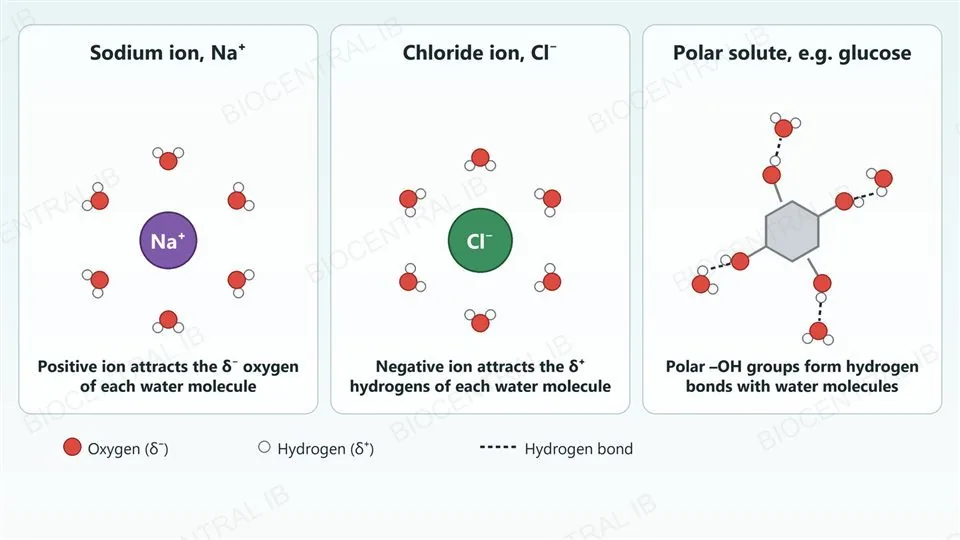
Three panels showing water molecules around dissolved particles: a sodium ion surrounded by six water molecules with their red oxygen atoms facing the ion, a chloride ion surrounded by six water molecules with their white hydrogen atoms facing the ion, and a polar solute with four hydroxyl groups joined to nearby water molecules by dashed hydrogen bonds, with a legend for oxygen, hydrogen and hydrogen bond.
D2.3.2

Water moves toward the more concentrated solution

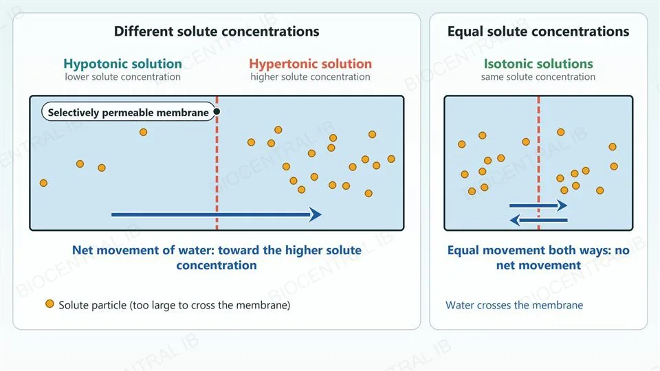
Osmosis is the net movement of water across a selectively permeable membrane from a solution with a lower solute concentration to one with a higher solute concentration.
  • Always describe the direction in terms of solute concentration, never water concentration.
  • Hypertonic means having a higher solute concentration than the solution it is compared with, hypotonic means lower, and isotonic means equal.
  • These terms are always comparative: a solution is hypertonic, hypotonic or isotonic only relative to another solution, such as a cell’s cytoplasm.
Why it mattersIn net terms, water always moves from the hypotonic solution into the hypertonic one.
A chamber split by a dashed membrane with a hypotonic solution on the left holding four solute particles and a hypertonic solution on the right holding eighteen, and a long arrow showing net movement of water toward the higher solute concentration; beside it, an isotonic chamber with nine particles on each side and two equal opposite arrows meaning no net movement.
D2.3.3

Predicting net water movement for a cell

The direction of net water movement into or out of a cell depends on comparing the solute concentration outside the cell with the concentration inside it.
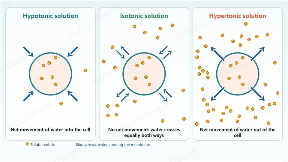
  • In a hypotonic environment there is net movement of water into the cell.
  • In a hypertonic environment there is net movement of water out of the cell.
  • In an isotonic environment water molecules still cross the membrane in both directions at equal rates: this is a dynamic equilibrium, not an absence of movement.
Why it mattersOnly the net movement is zero in an isotonic solution; individual water molecules keep moving.
Three identical cells each containing six solute particles: in a hypotonic solution with two particles outside, four inward arrows show net movement of water into the cell; in an isotonic solution with sixteen particles outside, pairs of equal in and out arrows show no net movement; in a hypertonic solution with forty particles outside, four outward arrows show net movement of water out of the cell.
Part two

Osmosis in tissues, cells and medicine

D2.3.4 – D2.3.7
D2.3.4

Plant tissue gains or loses water in solutions

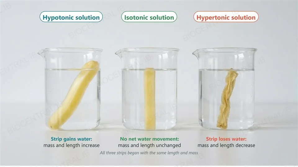
Plant tissue bathed in a hypotonic solution gains water by osmosis, while tissue bathed in a hypertonic solution loses water.
  • Tissue such as potato strips in a hypotonic solution takes in water, so its mass and length increase.
  • In a hypertonic solution the tissue loses water, so its mass and length decrease.
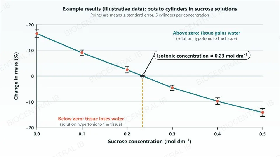
  • Measuring these changes across a range of external solute concentrations lets you find the concentration at which there is no net change.
Why it mattersUsing percentage change lets samples with different starting masses or lengths be compared fairly.
Three identical glass beakers, each with one potato strip: in the hypotonic solution the strip is swollen and longer and is labelled as gaining water, in the isotonic solution it is straight and unchanged, and in the hypertonic solution it is shrunken and wrinkled and is labelled as losing water.
D2.3.4

Reading the data to find the isotonic concentration

Plotting percentage change in mass or length against solute concentration gives a line, and the point where it crosses zero change is the isotonic concentration of the tissue.
  • At that concentration the external solution matches the tissue’s own solute concentration, so there is no net water movement.
  • Standard deviation describes the spread of repeated measurements around each mean, and standard error shows how precisely each mean is known.
  • Standard error can be drawn as error bars, and the formulae for these statistics do not have to be memorised.
Why it mattersOverlapping error bars suggest that a difference between two concentrations may not be reliable.
An example line graph with illustrative data: percentage change in mass of potato cylinders falls from about plus sixteen at zero sucrose concentration to about minus fourteen at 0.5 mol per cubic decimetre, with error bars on every point, crossing zero at an isotonic concentration of about 0.23 mol per cubic decimetre, with regions above and below zero labelled as gaining and losing water.
D2.3.5

Cells without a wall swell, burst or shrink

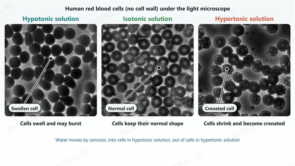
Animal cells have no cell wall, so osmosis changes their volume directly.
  • In a hypotonic medium the cell takes up water, swells and may burst; in red blood cells this bursting is called haemolysis.
  • In a hypertonic medium the cell loses water and shrinks, and its surface becomes shrivelled and spiky, which is called crenation.
  • In an isotonic medium the cell keeps its normal shape.
Why it mattersWith no wall to resist swelling, a cell’s volume depends entirely on the solute concentration outside it.
Three real light micrographs of human red blood cells: in a hypotonic solution the cells look swollen and rounded, in an isotonic solution they keep their normal shape, and in a hypertonic solution they are shrunken with a spiky, crenated surface; a leader in each panel points to a swollen, a normal and a crenated cell.
Zephyris · CC BY-SA 3.0 · Wikimedia Commons
D2.3.5

Contractile vacuoles bail the water out

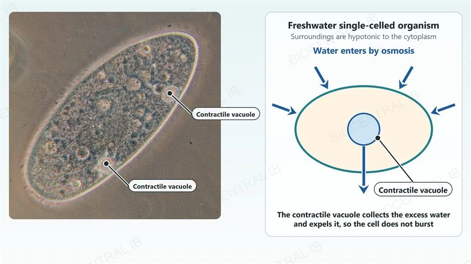
Freshwater unicellular organisms, such as Paramecium, live in a hypotonic environment and constantly gain water by osmosis.
  • Without a cell wall to resist swelling, they would burst if the water were not removed.
  • A contractile vacuole collects the excess water and expels it from the cell.
  • Removing the water is an active process that needs energy.
Why it mattersThe vacuole solves the same problem that a multicellular animal solves with isotonic tissue fluid, on the next slide.
A real micrograph of a Paramecium with two labelled contractile vacuoles, beside a diagram of a freshwater single-celled organism with blue arrows showing water entering by osmosis and a blue arrow carrying water out from the contractile vacuole.
Josh Grosse / Hämbörger · CC BY-SA 3.0 · Wikimedia
D2.3.5

Multicellular animals keep their tissue fluid isotonic

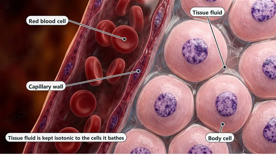
In a multicellular organism the cells are bathed in tissue fluid, whose solute concentration must be kept isotonic to the cells to prevent harmful changes.
  • If tissue fluid became hypotonic, cells without walls would take up water and swell.
  • If it became hypertonic, they would lose water and shrink.
  • Blood plasma and tissue fluid are held at a controlled solute concentration, a whole-organism answer to the problem a freshwater cell solves with a contractile vacuole.
Why it mattersCells without walls have no structural way to resist osmotic swelling or shrinkage, so regulation is essential.
A cross-section of tissue showing a blood capillary with red blood cells on the left and rounded pink body cells on the right, with clear tissue fluid in the gaps between them; labels point to a red blood cell, the capillary wall, the tissue fluid and a body cell, and a caption says tissue fluid is kept isotonic to the cells it bathes.
D2.3.6

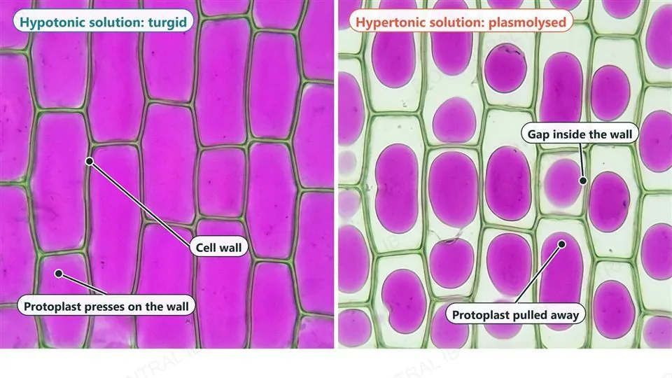
Cell walls turn water uptake into turgor

Plant cells have a cell wall, so osmosis changes the pressure inside the cell instead of letting it burst.
  • In a hypotonic medium water enters and the protoplast (membrane and cytoplasm) presses against the wall, building turgor pressure that makes the cell firm.
  • In a hypertonic medium water leaves and the protoplast shrinks away from the wall, which is called plasmolysis.
  • Plasmolysis is reversible if the cell returns to a hypotonic solution before its membrane is damaged.
Why it mattersTurgor pressure in many cells together keeps non-woody stems and leaves rigid.
Two microscope-style panels of rectangular plant cells: on the left, in a hypotonic solution, purple protoplasts fill every cell and press on the green cell walls; on the right, in a hypertonic solution, the walls are unchanged but each purple protoplast has shrunk into a rounded blob leaving a clear gap; labels point to a cell wall, a protoplast pressing on the wall, a gap inside the wall and a protoplast pulled away.
D2.3.7

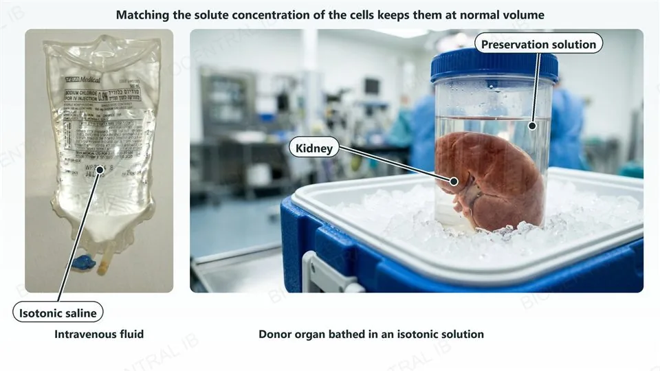
Isotonic solutions protect cells in medicine

A solution isotonic to human cells causes no net water movement, so it can be used to keep cells at their normal volume.
  • Intravenous fluids given in medical treatment, such as 0.9% saline, are isotonic to blood plasma so that red blood cells neither swell nor shrink.
  • Organs are bathed in an isotonic solution before transplantation to prevent osmotic damage to their cells.
  • A hypotonic or hypertonic fluid given by mistake would risk haemolysis or crenation of blood cells.
Why it mattersBoth uses rely on one principle: matching the external solute concentration to the internal one keeps cells intact.
On the left, a real photograph of a bag of 0.9 percent sodium chloride intravenous fluid labelled as isotonic saline; on the right, a photograph of a human kidney in a clear container of preservation solution packed in ice in a transport cooler, with leaders labelling the kidney and the preservation solution.
IV bag: Matanya (uploader) · CC BY-SA 3.0 · Wikimedia
Quick check

A red blood cell is placed in a solution with a lower solute concentration than its cytoplasm. Which prediction is correct?

Net movement of water out of the cell, so it shrinks and becomes crenated
Net movement of water into the cell, so it swells and may burst
No water movement at all, because the membrane is selectively permeable
Water moves toward the lower solute concentration, so the cell is unchanged
Correct answer: net movement of water into the cell, so it swells and may burst. The solution is hypotonic to the cell, and water moves toward the higher solute concentration, which is inside the cell (D2.3.2, D2.3.3). A red blood cell has no wall to resist swelling, so it may burst (haemolysis, D2.3.5).

Key vocabulary — SL core

D2.3.1 – D2.3.7: worth being able to define each in a sentence

Solvation
The dissolving of a solute in water, as water molecules gather around it by hydrogen bonding and attraction between charges.
Osmosis
The net movement of water across a selectively permeable membrane from lower to higher solute concentration.
Hypertonic / hypotonic / isotonic
Comparative terms for a solution with a higher, a lower, or an equal solute concentration than another solution.
Dynamic equilibrium
The state in an isotonic solution where water crosses the membrane both ways at equal rates, so there is no net movement.
Haemolysis
The bursting of red blood cells after they take up too much water in a hypotonic solution.
Crenation
The shrivelled, spiky appearance of an animal cell that has lost water in a hypertonic solution.
Contractile vacuole
An organelle in freshwater single-celled organisms that collects excess water and expels it from the cell.
Turgor pressure
The pressure that builds up when the protoplast of a walled cell presses against the cell wall in a hypotonic solution.
Plasmolysis
The shrinking of a walled cell’s protoplast away from its cell wall in a hypertonic solution.
Part three · HL

HL — water potential

D2.3.8 – D2.3.11
D2.3.8 · HL

Water potential: potential energy per unit volume

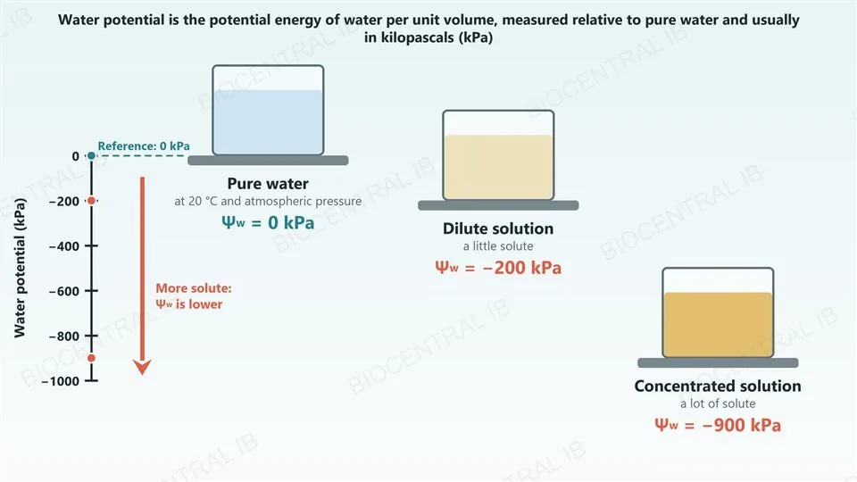
Water potential (Ψw) is the potential energy of water per unit volume.
  • The absolute potential energy of water cannot be measured, so values are given relative to pure water at atmospheric pressure and 20 °C.
  • Pure water under those conditions is assigned a water potential of 0 kPa, and the units are usually kilopascals (kPa).
  • Dissolving a solute lowers the water potential below zero, so most biological solutions have a negative Ψw.
Why it mattersA more negative Ψw means water has less potential energy per unit volume.
A vertical scale of water potential in kilopascals from 0 down to minus 1000, beside three beakers on shelves at matching heights: pure water at 0 kPa marked as the reference, a dilute solution at minus 200 kPa and a concentrated solution at minus 900 kPa, with a downward arrow labelled that more solute means a lower water potential.
D2.3.9 · HL

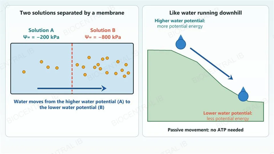
Water moves from higher to lower water potential

Water moves from a region of higher water potential to a region of lower water potential.
  • The reason is potential energy: like any object, water tends to move toward a lower energy state.
  • This is the same direction as in D2.3.2, where it was described using solute concentration.
  • The movement is passive and needs no ATP.
  • Net movement continues until the water potentials are equal, or until an opposing pressure stops it.
Why it mattersOne rule lets you predict osmosis in any situation, including walled plant cells.
Two solutions separated by a membrane, solution A at minus 200 kPa with four solute particles and solution B at minus 800 kPa with sixteen, with an arrow showing water moving from A to B; beside it, a droplet at the top of a slope labelled higher water potential and more potential energy rolling to the bottom labelled lower water potential and less potential energy, captioned that the movement is passive with no ATP needed.
D2.3.10 · HL

Solute and pressure potentials add up in walled cells

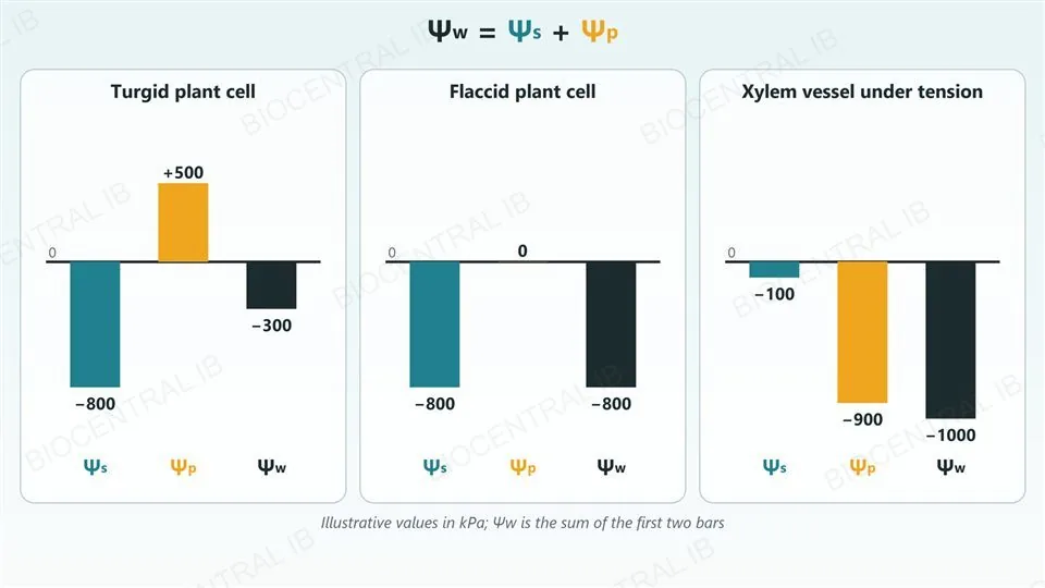
For a cell with a wall, water potential is the sum of solute potential and pressure potential: Ψw = Ψs + Ψp.
  • Solute potential (Ψs) is zero or negative: dissolved solutes only lower it, and more solute makes it more negative.
  • Pressure potential (Ψp) is generally positive inside cells, because the wall pushes back on the protoplast.
  • Negative pressure potentials occur in xylem vessels, where sap is transported under tension.
Why it mattersIn a turgid cell a positive Ψp raises Ψw toward zero; in a flaccid cell Ψp is zero, so Ψw equals Ψs.
The equation water potential equals solute potential plus pressure potential above three bar charts in kilopascals with illustrative values: a turgid plant cell with solute potential minus 800, pressure potential plus 500 and water potential minus 300; a flaccid plant cell with minus 800, 0 and minus 800; and a xylem vessel under tension with minus 100, minus 900 and minus 1000.
D2.3.11 · HL

Potentials explain how plant tissue responds

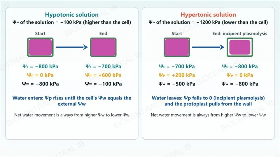
The changes in plant tissue bathed in a hypotonic or hypertonic solution can be explained in terms of solute and pressure potentials.
  • In a hypotonic solution the external Ψw is higher, so water enters; Ψp rises as the protoplast presses on the wall until the cell’s Ψw equals the external Ψw and the cell is turgid.
  • In a hypertonic solution the external Ψw is lower, so water leaves and Ψp falls to zero as the protoplast pulls away from the wall (incipient plasmolysis).
  • At incipient plasmolysis Ψw equals Ψs alone, which is used to estimate a cell’s solute potential.
Why it mattersThe rule never changes: net movement is always from higher Ψw to lower Ψw.
Two panels with illustrative values: in a hypotonic solution of water potential minus 100 kPa a plant cell goes from solute potential minus 800, pressure potential 0 and water potential minus 800 to minus 700, plus 600 and minus 100, with the protoplast filling the wall; in a hypertonic solution of minus 1200 kPa a cell goes from minus 700, plus 200 and minus 500 to minus 800, 0 and minus 800 at incipient plasmolysis, with the protoplast shrunk from the wall.
Quick check · HL

A flaccid plant cell with Ψs = −800 kPa and Ψp = 0 kPa is placed in a solution with Ψw = −100 kPa. What happens?

There is no net movement, because Ψp is zero
Water enters, because the cell’s Ψw (−800 kPa) is lower than the solution’s, and Ψp rises until the cell’s Ψw reaches −100 kPa
Water leaves the cell, because the solution has the higher water potential
Water enters until Ψs reaches 0 kPa, when the cell is turgid
Correct answer: water enters, and Ψp rises until the cell’s Ψw reaches −100 kPa. Ψw = Ψs + Ψp = −800 + 0 = −800 kPa, which is lower than −100 kPa, so water moves in (D2.3.9). As the protoplast presses on the wall Ψp rises, and net movement stops when the cell’s Ψw equals −100 kPa (D2.3.11). Solute potential can never be positive, so it cannot reach 0 kPa in a solution.

Key vocabulary — water potential · HL

HL only: D2.3.8 – D2.3.11

Water potential (Ψw)
The potential energy of water per unit volume, relative to pure water at atmospheric pressure and 20 °C, which is 0 kPa; water moves from higher to lower Ψw.
Solute potential (Ψs)
The part of water potential due to dissolved solutes; always zero or negative.
Pressure potential (Ψp)
The part of water potential due to pressure; generally positive in walled cells, negative in xylem vessels under tension.
Turgid cell
A walled cell with a positive Ψp, pressed firmly against its wall; its Ψw is closer to zero than its Ψs.
Flaccid cell
A walled cell with no turgor pressure: Ψp is zero, so Ψw equals Ψs.
Incipient plasmolysis
The point at which the protoplast just begins to pull away from the wall: Ψp is zero, so Ψw equals Ψs.

Where this shows up again

A1.1 · Water
Solvation (D2.3.1) rests on the hydrogen bonds and polarity of water met in A1.1.2 and on its solvent properties in A1.1.5. Explain why a polar molecule dissolves in water but a hydrocarbon does not.
B2.1 · Membranes
B2.1.5 explains osmosis in terms of random particle movement, impermeability to solutes and aquaporins. Outline how a selectively permeable membrane makes net water movement depend on solute concentration.
B3.2 · Transport · HL link
Tissue fluid (B3.2.11 and B3.2.12) is the fluid kept isotonic to cells in D2.3.5, and the negative pressure potential in xylem (D2.3.10) links to transpiration (B3.2.7). Explain why negative Ψp helps draw water up a plant.
D2.3 · Linking questions from the guide
What variables influence the direction of movement of materials in tissues? What are the implications of solubility differences between chemical substances for living organisms?

D2.3 Water potential — one-page recap

Screenshot this slide to revise from

Water as the solvent
  • Polar water surrounds ions (δ⁻ oxygen to cations, δ⁺ hydrogen to anions) and forms hydrogen bonds with polar solutes.
Osmosis and tonicity
  • Water moves to the higher solute concentration. Hypertonic higher, hypotonic lower, isotonic equal, always comparative.
Cells without a wall
  • Hypotonic: swell and may burst (haemolysis). Hypertonic: shrink and crenate. Freshwater cells use a contractile vacuole; animals keep isotonic tissue fluid.
Cells with a wall
  • Hypotonic: turgor pressure builds. Hypertonic: plasmolysis. Tissue data give the isotonic concentration where change is zero; error bars show standard error.
Medicine
  • 0.9% saline IV fluids and organ-bathing solutions are isotonic, so cells keep their normal volume.
HL · Water potential
  • Ψw in kPa, pure water 0. Water moves from higher to lower Ψw. Ψw = Ψs + Ψp; Ψs ≤ 0, Ψp positive in cells, negative in xylem.

Water follows the solutes, and the energy

Wherever solute concentration differs across a membrane, water moves toward the more concentrated side. Whether that swells a cell, bursts it, firms it up or shrinks it away from its wall depends only on what stands in the way: nothing, a contractile vacuole, a controlled tissue fluid, or a wall. At HL, the same story is told with potential energy.
D2.3 Water potential · BioCentral IB
Use ↓ ↑ or click to navigate
01 / 20