BioCentral IBBioCentral IB
IB Biology · Theme D · D3.3

Homeostasis

Homeostasis is the maintenance of a stable internal environment while the outside world changes. This topic follows the negative feedback loops that hold blood glucose and body temperature steady, and at HL goes on to the kidney’s control of the blood’s osmotic concentration and the redirection of blood to the organs that are most active.
Guiding questions

How are constant internal conditions maintained in humans?

What are the benefits to organisms of maintaining constant internal conditions?

Part one

Keeping conditions constant

D3.3.1 – D3.3.2
D3.3.1

The internal environment is held within preset limits

Homeostasis is the maintenance of the internal environment within preset limits, despite fluctuations in the external environment.
  • Each variable has a set point, and a corrective response starts when its value drifts outside the limits.
  • In humans the variables to know are body temperature, blood pH, blood glucose concentration and blood osmotic concentration.
  • Body temperature is about 37 °C, blood pH about 7.35 to 7.45, blood glucose about 4 to 6 mmol dm−3 and blood osmotic concentration about 280 to 300 mOsm L−1.
  • Enzymes and other cell processes work only in a narrow range of conditions, so large swings would damage cells.
Why it mattersConstant internal conditions let cells work reliably even when the outside world changes.
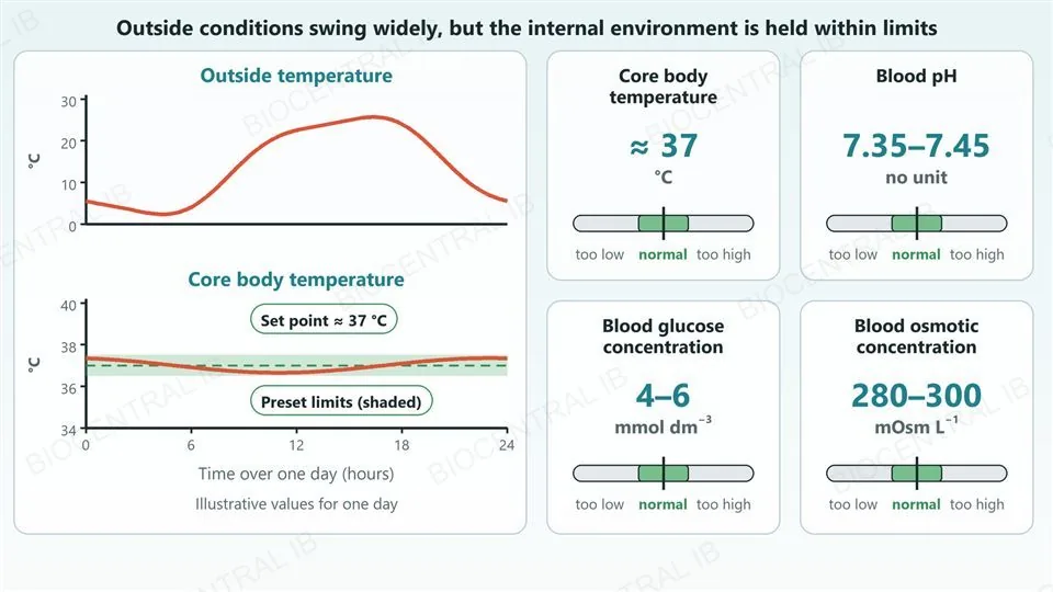
Left, two graphs over one day. The outside temperature rises and falls by more than 20 degrees Celsius, while the core body temperature stays in a narrow shaded band around a dashed set point of about 37 degrees Celsius. Right, four cards showing the normal value of each homeostatic variable: core body temperature about 37 degrees Celsius, blood pH 7.35 to 7.45, blood glucose concentration 4 to 6 millimoles per cubic decimetre, and blood osmotic concentration 280 to 300 milliosmoles per litre, each with a gauge marked too low, normal and too high.
D3.3.2

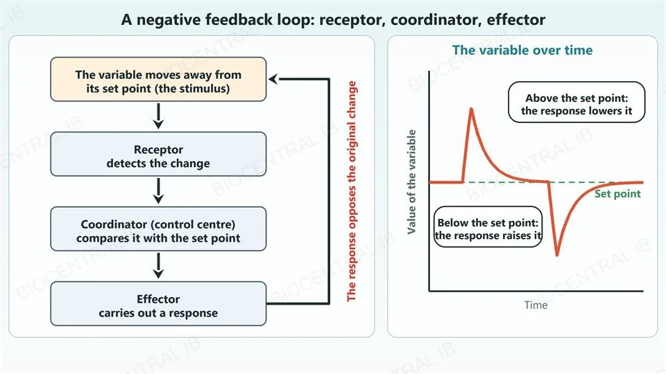
A negative feedback loop has a receptor, a coordinator and an effector

In negative feedback the response reverses the change that triggered it, which returns the variable to its set point.
  • A receptor detects the value of the variable, and a coordinator (control centre) compares it with the set point.
  • An effector then carries out a response that opposes the change.
  • The loop works from both sides: a rise above the set point triggers a response that lowers the variable, and a fall below it triggers a response that raises it.
Why it mattersThe same loop design is used for glucose, temperature and water balance.
Left, a flow chart of boxes joined by arrows: the variable moves away from its set point (the stimulus), the receptor detects the change, the coordinator (control centre) compares it with the set point, and the effector carries out a response; a return arrow labelled that the response opposes the original change leads back to the first box. Right, a graph of the value of a variable over time with a dashed set point line: it rises above the set point and the response lowers it back, then it falls below the set point and the response raises it back.
D3.3.2

Negative feedback keeps a system stable

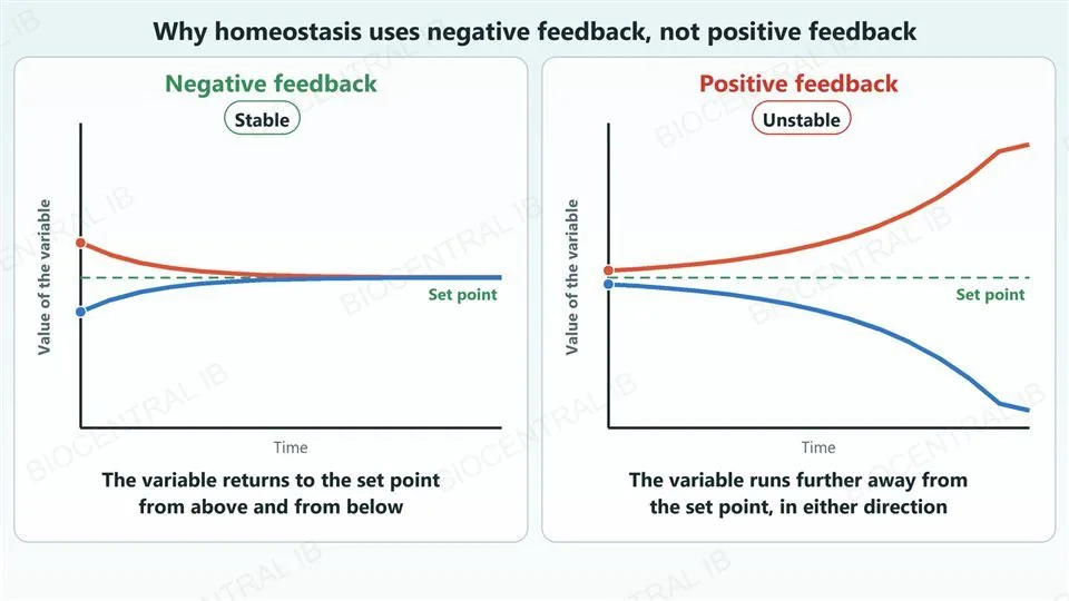
Homeostasis uses negative feedback because only negative feedback keeps a variable near its set point.
  • Negative feedback returns a variable to the set point from values above it and from values below it.
  • Positive feedback amplifies a change, so the variable moves further away from the set point, whichever direction it started in.
  • A system controlled by positive feedback would run away to extreme values, so it could not hold a constant internal state.
Why it mattersA control system that holds a constant state must oppose change, not amplify it.
Two graphs side by side, each with a dashed set point line and two curves that start slightly above and slightly below it. In the negative feedback graph, labelled stable, both curves return to the set point. In the positive feedback graph, labelled unstable, the curve that started above climbs steadily away upward and the curve that started below falls steadily away downward.
Part two

Blood glucose and diabetes

D3.3.3 – D3.3.4
D3.3.3

Two kinds of islet cell make antagonistic hormones

Blood glucose is regulated by hormones made by endocrine cells in the pancreas.
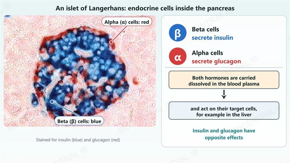
  • Clusters of endocrine cells called islets of Langerhans contain beta (β) cells and alpha (α) cells.
  • β cells secrete insulin and α cells secrete glucagon, and the concentration of glucose in the blood controls how much of each is secreted.
  • Both hormones pass into the blood and are carried in the plasma to their target cells, mainly in the liver and muscle.
Why it mattersThe same organ makes both hormones, so it can push blood glucose in either direction.
A real micrograph of one human pancreatic islet of Langerhans, stained for insulin in blue and glucagon in red. The blue beta cells fill most of the islet and the red alpha cells lie mainly around its edge; two labels with leader dots point at a blue area and a red area. Right, cards saying that beta cells secrete insulin, alpha cells secrete glucagon, both hormones are carried dissolved in the blood plasma and act on target cells such as the liver, and that insulin and glucagon have opposite effects.
Photo: Afferent, CC BY-SA 3.0 · Wikimedia Commons
D3.3.3

Insulin lowers blood glucose and glucagon raises it

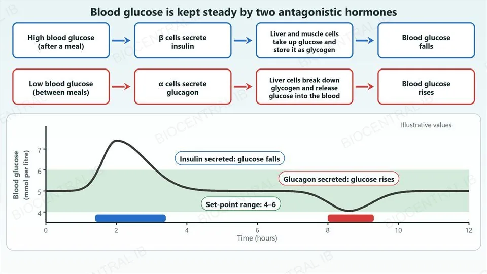
The two hormones form a negative feedback loop that keeps blood glucose within its set-point range.
  • When blood glucose is high, β cells secrete insulin, and liver and muscle cells take up glucose and store it as glycogen.
  • When blood glucose is low, α cells secrete glucagon, and liver cells break down glycogen and release glucose into the blood.
  • Each response removes its own stimulus, so blood glucose returns to the range of about 4 to 6 mmol dm−3.
Why it mattersInsulin lowers and glucagon raises: reversing them is the most common exam error.
Top, two rows of four boxes joined by arrows. Blue row: high blood glucose after a meal, β cells secrete insulin, liver and muscle cells take up glucose and store it as glycogen, blood glucose falls. Red row: low blood glucose between meals, α cells secrete glucagon, liver cells break down glycogen and release glucose into the blood, blood glucose rises. Bottom, an illustrative graph of blood glucose over twelve hours inside a shaded set-point range of 4 to 6: it rises to about 7.4 after a meal while insulin is secreted, then returns to about 5, and later dips to about 4 while glucagon is secreted.
D3.3.4

Type 1 and type 2 diabetes have different causes

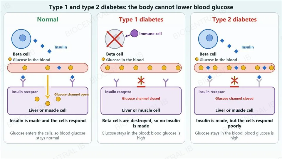
In both types of diabetes the body cannot bring blood glucose down, so it stays high.
  • In type 1 diabetes the immune system destroys the β cells, so little or no insulin is made.
  • In type 2 diabetes the target cells respond poorly to insulin, which is called insulin resistance, even though insulin is present.
  • Either way glucose stays in the blood instead of entering the cells, but the two types need different treatments.
Why it mattersType 1 is a problem of insulin supply, and type 2 is a problem of the response to insulin.
Three columns compared. Normal: a beta cell releases insulin, which binds receptors on a liver or muscle cell, the glucose channel opens and glucose enters the cell, so blood glucose stays normal. Type 1 diabetes: an immune cell attacks the beta cell, which is crossed out, so no insulin is made, the receptors are empty, the glucose channel is closed and glucose stays in the blood. Type 2 diabetes: the beta cell makes insulin and it binds to faded receptors, but the glucose channel stays closed because the cells respond poorly, so glucose stays in the blood.
D3.3.4

Risk factors, prevention and treatment differ

The two types differ in their risk factors and in how they are prevented and treated.
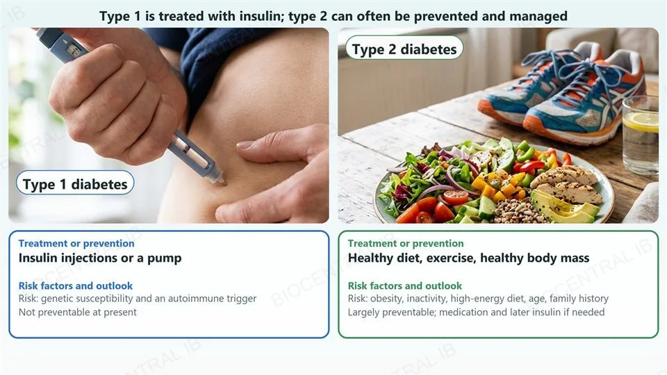
  • Type 1 has a genetic susceptibility and an autoimmune trigger, and it cannot be prevented at present; it is treated with insulin injections or an insulin pump.
  • Risk factors for type 2 include obesity, physical inactivity, a high-energy diet, age and family history.
  • Type 2 is largely preventable, and it is managed with a healthy diet, exercise and a healthy body mass, with medication and later insulin if needed.
Why it mattersLifestyle changes can prevent or delay many cases of type 2 diabetes.
Two photographs. Left, headed type 1 diabetes: a hand pressing an insulin pen against the skin of an abdomen. Right, headed type 2 diabetes: a plate of salad, vegetables, grilled chicken, avocado and whole grains on a wooden table beside running shoes and a glass of water. Under each photograph a card gives the treatment or prevention and the risk factors: type 1 is treated with insulin injections or a pump, its risk is genetic susceptibility and an autoimmune trigger, and it is not preventable at present; type 2 is prevented and managed with a healthy diet, exercise and a healthy body mass, its risks are obesity, inactivity, a high-energy diet, age and family history.
Part three

Thermoregulation

D3.3.5 – D3.3.6
D3.3.5

Body temperature is controlled by negative feedback

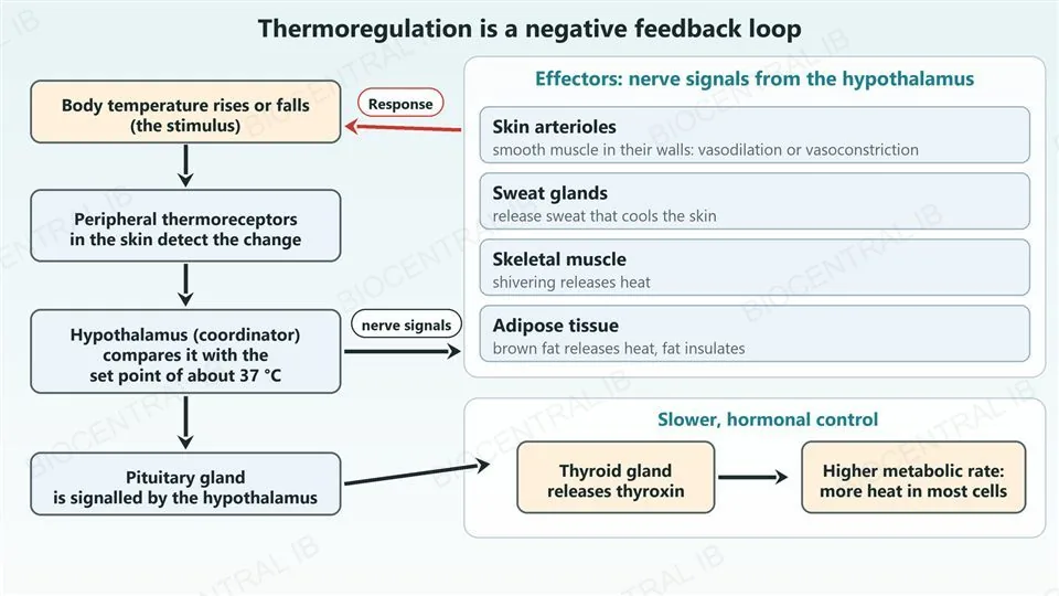
Thermoregulation uses receptors, a coordinator in the brain and several effectors to hold body temperature near a set point of about 37 °C.
  • Peripheral thermoreceptors in the skin detect changes in temperature and send signals to the brain.
  • The hypothalamus compares the signals with the set point and sends nerve signals to the effectors.
  • The hypothalamus also acts on the pituitary gland, which leads to secretion of thyroxin; thyroxin raises the metabolic rate, so more heat is made.
  • Effectors include skeletal muscle, adipose tissue, sweat glands and the smooth muscle of skin arterioles.
Why it mattersNerve signals give a fast response, and thyroxin gives a slower, longer-lasting one.
A flow chart. On the left, boxes joined by downward arrows: body temperature rises or falls (the stimulus), peripheral thermoreceptors in the skin detect the change, the hypothalamus (coordinator) compares it with the set point of about 37 degrees Celsius, and the pituitary gland is signalled by the hypothalamus. From the hypothalamus an arrow labelled nerve signals leads to a card listing four effectors: skin arterioles (smooth muscle, vasodilation or vasoconstriction), sweat glands (release sweat), skeletal muscle (shivering) and adipose tissue (brown fat releases heat, fat insulates). A red arrow labelled response leads from the effectors back to the stimulus. From the pituitary gland an arrow leads to the thyroid gland releasing thyroxin, then to a higher metabolic rate and more heat in most cells.
D3.3.6

When the body is too hot, it loses heat

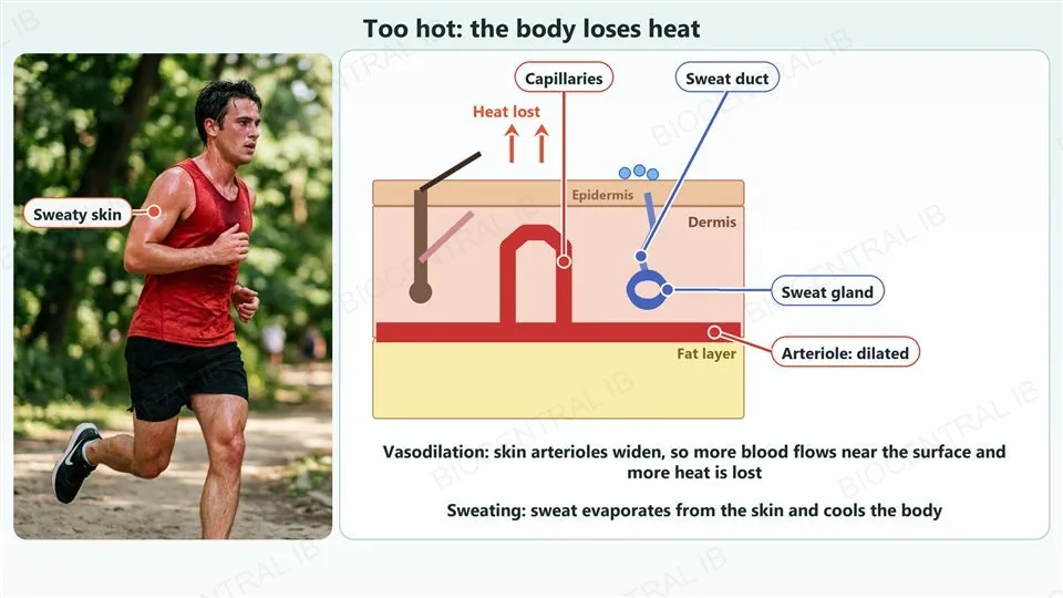
Birds and mammals control body temperature by physiological and behavioural means, and in humans two physiological responses cool the body.
  • Vasodilation: the arterioles in the skin widen, so more blood flows near the surface and more heat is lost to the surroundings.
  • Sweating: sweat glands release sweat, and its evaporation from the skin cools the body.
  • Behaviour helps too, for example moving into the shade.
Why it mattersVasodilation means heat loss.
Left, a photograph of a runner on a hot day in a park, with sweat on the skin, and a label with a leader dot pointing at the sweaty skin of the shoulder. Right, a labelled cross-section of skin during heat. A wide, dilated arteriole runs along the base of the dermis and a loop of capillaries rises to just under the surface; a sweat gland with a duct opens onto the surface where drops of sweat sit; a hair lies flat. Labels with leader dots name the capillaries, the sweat duct, the sweat gland and the dilated arteriole, and red arrows show heat lost from the skin. Text says that vasodilation means more blood flows near the surface and more heat is lost, and that sweat evaporating cools the body.
D3.3.6

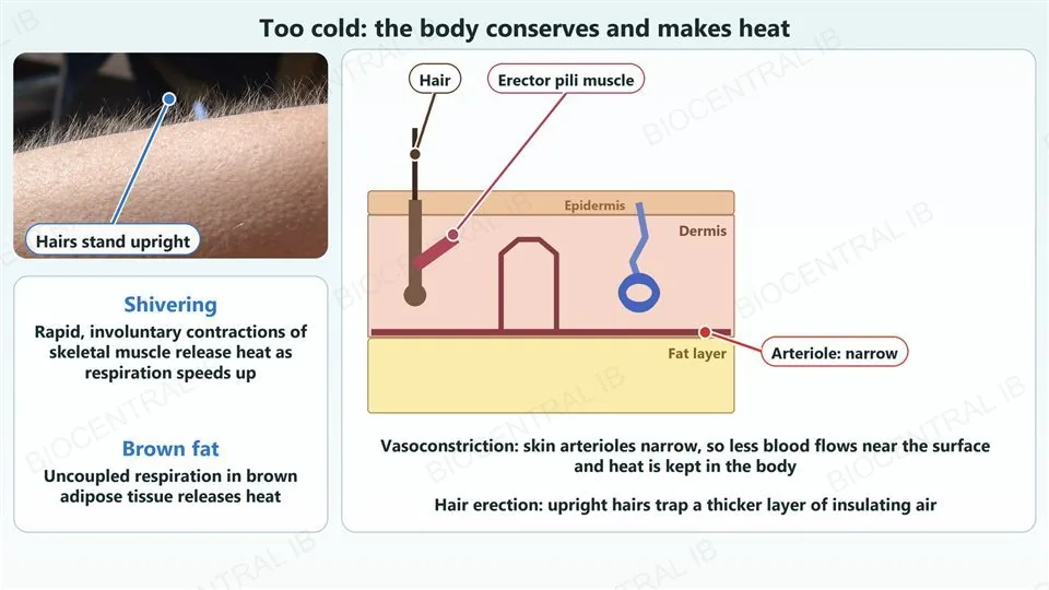
When the body is too cold, it keeps and makes heat

In the cold, skin responses reduce heat loss and muscle activity makes extra heat.
  • Vasoconstriction: skin arterioles narrow, so less blood flows near the surface and heat is kept in the body.
  • Hair erection: the erector muscles pull hairs upright, trapping a thicker layer of insulating air next to the skin.
  • Shivering: rapid, involuntary contractions of skeletal muscle release heat.
Why it mattersVasoconstriction means heat kept in, so do not swap it with vasodilation.
Left, a real close-up photograph of a forearm with goose bumps, the fine hairs standing upright, with a label and leader dot pointing at the upright hairs, and a card noting that shivering releases heat and that uncoupled respiration in brown adipose tissue releases heat. Right, a labelled cross-section of skin in the cold. The arteriole along the base of the dermis is narrow, the hair stands upright, and the erector pili muscle attached to the hair follicle is contracted and thick. Labels with leader dots name the hair, the erector pili muscle and the narrow arteriole. Text says vasoconstriction means less blood flows near the surface and heat is kept, and upright hairs trap insulating air.
Photo: EverJean, CC BY 2.0 · Wikimedia Commons
D3.3.6

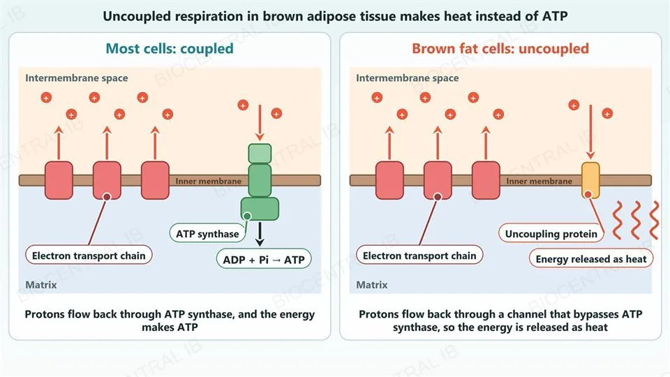
Brown adipose tissue makes heat by uncoupled respiration

Brown adipose tissue releases the energy of respiration directly as heat instead of using it to make ATP.
  • In most cells the electron transport chain pumps protons across the inner mitochondrial membrane, and the protons flow back through ATP synthase, which makes ATP.
  • In brown fat cells the protons flow back through a channel protein instead, so respiration is uncoupled from ATP production.
  • The energy of the proton gradient is then released as heat, which warms the body.
Why it mattersMaking heat is the job of brown fat, not a by-product.
Two diagrams of a mitochondrial inner membrane, with the intermembrane space above and the matrix below. Left, most cells, coupled: three red electron transport chain complexes pump protons up into the intermembrane space, and the protons flow back down through ATP synthase, where ADP and phosphate become ATP. Right, brown fat cells, uncoupled: the same three complexes pump protons up, but the protons flow back down through an uncoupling protein channel instead of ATP synthase, and wavy red lines show the energy released as heat.
Quick check

A person walks out into freezing weather. Which set of responses conserves and makes heat?

Vasoconstriction of skin arterioles, sweating and hair erection
Vasoconstriction of skin arterioles, hair erection and shivering
Vasodilation of skin arterioles, hair erection and less muscle activity
Vasodilation of skin arterioles, sweating and shivering
Correct answer: vasoconstriction of skin arterioles, hair erection and shivering. Narrow skin arterioles carry less blood to the surface, so less heat is lost; upright hairs trap insulating air; and shivering muscles release heat. Vasodilation and sweating both increase heat loss, so they belong to the hot response (D3.3.6).

Key vocabulary — homeostasis and blood glucose

D3.3.1 – D3.3.4: worth being able to define each in a sentence

Homeostasis
Maintenance of the internal environment within preset limits, despite changes outside.
Set point
The target value of a variable that the body works to maintain.
Negative feedback
A response that reverses the change that triggered it, returning the variable to its set point.
Positive feedback
A response that amplifies the change that triggered it, moving the variable further away.
Islets of Langerhans
Clusters of endocrine cells in the pancreas: β cells make insulin, α cells make glucagon.
Insulin
A hormone that lowers blood glucose by making cells take up glucose and store it as glycogen.
Glucagon
A hormone that raises blood glucose by making liver cells break down glycogen.
Type 1 diabetes
An autoimmune destruction of β cells, so little or no insulin is made.
Type 2 diabetes
A poor response of target cells to insulin (insulin resistance), so glucose stays in the blood.

Key vocabulary — thermoregulation

D3.3.5 – D3.3.6: worth being able to define each in a sentence

Thermoreceptor
A receptor that detects temperature; peripheral thermoreceptors are in the skin.
Hypothalamus
The part of the brain that compares body temperature with the set point.
Thyroxin
A hormone that raises the metabolic rate and so the heat production of cells.
Vasodilation
Widening of skin arterioles, so more heat is lost.
Vasoconstriction
Narrowing of skin arterioles, so less heat is lost.
Sweating
Release of sweat, which cools the skin as it evaporates.
Shivering
Rapid involuntary contractions of skeletal muscle that release heat.
Hair erection
Erector muscles pull hairs upright, trapping insulating air.
Uncoupled respiration
Protons return through a channel that bypasses ATP synthase, so energy is released as heat.
Part four · HL

HL — the kidney and blood supply

D3.3.7 – D3.3.11
D3.3.7 · HL

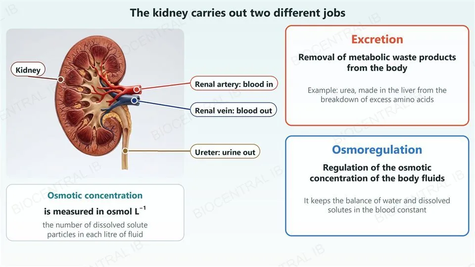
Excretion and osmoregulation are different jobs

The kidney carries out both excretion and osmoregulation, but they are not the same process.
  • Excretion is the removal of metabolic waste products from the body, for example urea.
  • Osmoregulation is the regulation of the osmotic concentration of the body fluids, so the balance of water and dissolved solutes in the blood stays constant.
  • Osmotic concentration is measured in osmoles per litre (osmol L−1).
Why it mattersRemoving urea is excretion, and controlling water balance is osmoregulation.
Left, a photograph of an anatomical model of a human kidney cut open, with labels and leader dots for the kidney, the renal artery (blood in), the renal vein (blood out) and the ureter (urine out), and a card saying osmotic concentration is measured in osmol per litre, the number of dissolved solute particles in each litre of fluid. Right, two cards: excretion is the removal of metabolic waste products from the body, for example urea made in the liver from the breakdown of excess amino acids; osmoregulation is the regulation of the osmotic concentration of the body fluids, keeping the balance of water and dissolved solutes in the blood constant.
D3.3.8–10 · HL

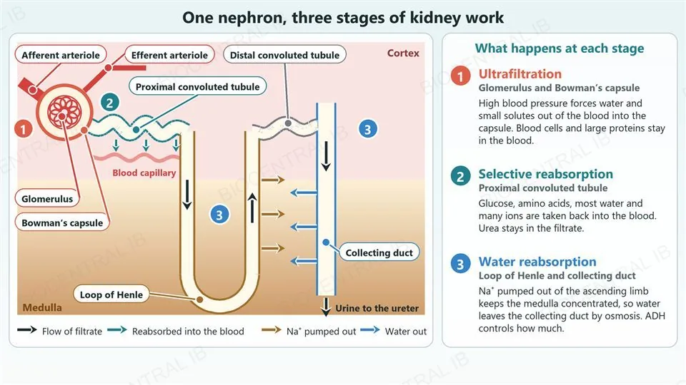
The nephron does its work in three stages

Each nephron filters the blood, keeps what the body needs and adjusts water loss in three stages.
  • Stage 1, ultrafiltration: blood pressure forces water and small solutes out of the glomerulus into Bowman’s capsule as filtrate.
  • Stage 2, selective reabsorption: the proximal convoluted tubule returns glucose, amino acids, most water and many ions to the blood.
  • Stage 3, water reabsorption: the loop of Henle pumps Na+ into the medulla, so water leaves the collecting duct by osmosis; ADH controls how much.
  • What remains in the collecting duct is urine.
Why it mattersThe next slides take one stage each and show how it happens.
A labelled diagram of one nephron. In the cortex, an afferent arteriole (wide) brings blood into a glomerulus inside Bowman’s capsule and a narrower efferent arteriole leaves; the proximal convoluted tubule leads from the capsule, with a blood capillary beside it and small arrows showing substances passing from the tubule into the blood. The tubule continues into the loop of Henle, which dips into the medulla and returns, with arrows showing Na+ pumped out of the ascending limb, then a distal convoluted tubule that joins the collecting duct. The collecting duct runs down through the medulla with arrows showing water leaving by osmosis, and urine leaves towards the ureter. Numbered badges mark three stages: 1 ultrafiltration at the glomerulus and Bowman’s capsule, 2 selective reabsorption in the proximal convoluted tubule, 3 water reabsorption at the loop of Henle and collecting duct. A panel on the right explains where and how each stage happens.
D3.3.8 · HL

Stage 1: ultrafiltration removes small solutes from the blood plasma

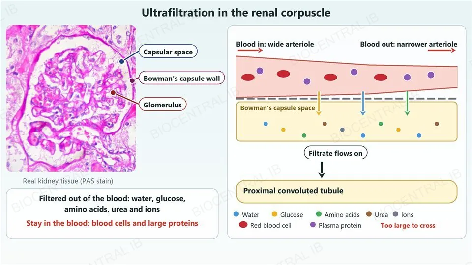
In the renal corpuscle, high blood pressure forces fluid out of the glomerulus and into Bowman’s capsule.
  • The glomerulus is a knot of capillaries, and the wall between it and Bowman’s capsule acts as a filter.
  • Water and small solutes such as glucose, amino acids, urea and ions pass into the capsule as filtrate.
  • Blood cells and large plasma proteins are too big to cross, so they stay in the blood.
  • The pressure is high because the arteriole leaving the glomerulus is narrower than the one entering it.
  • Filtration depends on size alone, so it is not selective.
Why it mattersThe filtrate still contains useful substances that must be recovered.
Left, a real micrograph of a renal corpuscle (PAS stain) with three labels and leader dots: the capsular space, the wall of Bowman’s capsule, and the glomerulus. Right, a diagram of a capillary running left to right, wide where blood comes in and narrower where it leaves, with red blood cells and purple plasma proteins staying inside it. Below its wall, coloured arrows show small molecules crossing into Bowman’s capsule space, where dots for water, glucose, amino acids, urea and ions collect; an arrow shows the filtrate flowing on into the proximal convoluted tubule. A card notes that water, glucose, amino acids, urea and ions are filtered out and that blood cells and large proteins stay in the blood.
Photo: Ed Uthman, CC BY-SA 2.0 · Wikimedia Commons
D3.3.8 · HL

Stage 2: the proximal convoluted tubule reabsorbs useful substances

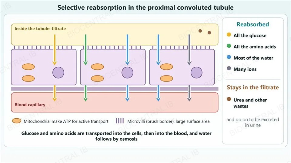
The filtrate flows from Bowman’s capsule into the proximal convoluted tubule, where useful substances are taken back into the blood.
  • The cells of the tubule have microvilli and many mitochondria, which give a large surface area and ATP for active transport.
  • Essentially all the glucose and amino acids are reabsorbed by active transport, together with many ions, and most of the water follows by osmosis.
  • Urea and other unwanted solutes stay in the filtrate and are excreted in urine.
Why it mattersReabsorption, not filtration, decides what the body keeps and what becomes waste.
A diagram of the wall of the proximal convoluted tubule. The filtrate in the tubule lumen is at the top, three tubule cells with microvilli, mitochondria and a nucleus are in the middle, and a blood capillary is at the bottom. Coloured arrows carry glucose, amino acids, water and ions from the lumen through the cells into the capillary. A card lists what is reabsorbed (all the glucose, all the amino acids, most of the water, many ions) and what stays in the filtrate (urea and other wastes, which go on to be excreted in urine). A key marks the mitochondria and the microvilli.
D3.3.9 · HL

Stage 3: the loop of Henle builds a concentrated medulla

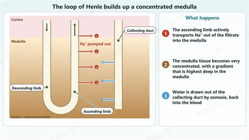
The ascending limb of the loop of Henle actively transports sodium ions out of the filtrate.
  • The sodium ions are pumped into the tissue of the renal medulla, which keeps its osmotic concentration high.
  • The medulla is therefore much more concentrated than the filtrate in the collecting duct.
  • As the collecting duct passes through the medulla, water can be drawn out of it by osmosis.
Why it mattersA concentrated medulla is what makes concentrated urine possible.
A diagram of a nephron in the kidney, with a pale cortex above a medulla that darkens with depth to show a rising osmotic concentration. A descending limb goes down into the medulla, turns in a hairpin, and returns as an ascending limb; red arrows show sodium ions pumped out of the ascending limb into the medulla. Beside them a collecting duct runs down through the medulla, with blue arrows showing water leaving it. Labels with leader dots name the descending limb, the ascending limb and the collecting duct. Right, three numbered steps: the ascending limb actively transports sodium ions out into the medulla, the medulla tissue becomes very concentrated with the highest concentration deep in the medulla, and water is drawn out of the collecting duct by osmosis back into the blood.
D3.3.10 · HL

Stage 3: osmoreceptors and ADH keep the blood’s osmotic concentration steady

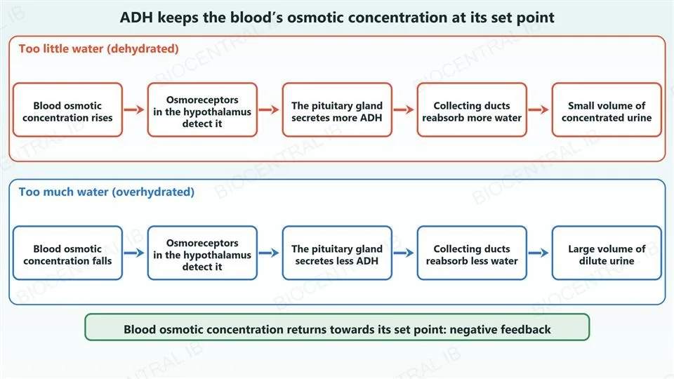
A negative feedback loop adjusts water reabsorption so that blood osmotic concentration stays at its set point.
  • Osmoreceptors in the hypothalamus monitor the osmotic concentration of the blood.
  • When it is high the pituitary gland secretes more antidiuretic hormone (ADH), and when it is low it secretes less.
  • ADH acts on the collecting ducts: more ADH means more water reabsorbed and a small volume of concentrated urine.
Why it mattersDehydration raises ADH, and overhydration lowers it.
Two rows of five boxes joined by arrows. Red row, too little water (dehydrated): blood osmotic concentration rises, osmoreceptors in the hypothalamus detect it, the pituitary gland secretes more ADH, collecting ducts reabsorb more water, small volume of concentrated urine. Blue row, too much water (overhydrated): blood osmotic concentration falls, osmoreceptors in the hypothalamus detect it, the pituitary gland secretes less ADH, collecting ducts reabsorb less water, large volume of dilute urine. A banner below says that blood osmotic concentration returns towards its set point, which is negative feedback.
D3.3.10 · HL

Stage 3: ADH switches aquaporins between vesicles and the membrane

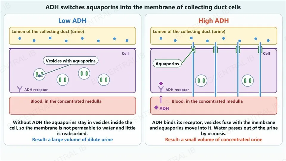
ADH changes how permeable collecting duct cells are to water by moving aquaporins, which are water channel proteins.
  • With high ADH, aquaporins move from vesicles inside the cell into the cell membrane.
  • The membrane is then permeable to water, so water is reabsorbed by osmosis into the concentrated medulla.
  • With low ADH, the aquaporins are removed from the membrane back into vesicles, less water is reabsorbed, and the urine is dilute.
Why it mattersADH does not make new channels each time; it moves existing ones.
Two panels of a collecting duct cell, with the urine in the lumen above, the cell in the middle and the blood in the concentrated medulla below. Left, low ADH: the green aquaporins sit inside four vesicles within the cell and none are in the membrane, so water stays in the urine; a label with a leader dot names the vesicles with aquaporins, and the result is a large volume of dilute urine. Right, high ADH: ADH from the blood binds a receptor on the cell, the vesicles fuse with the membrane so that aquaporins sit in the top membrane, and blue arrows show water passing from the lumen through the cell into the blood; a label with a leader dot names an aquaporin, and the result is a small volume of concentrated urine.
D3.3.11 · HL

Blood is redirected to the organs that are most active

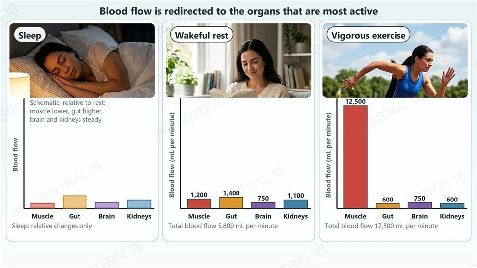
The share of the blood that flows to each organ changes with the body’s level of activity.
  • In vigorous exercise flow to skeletal muscle rises steeply, from about 1,200 to about 12,500 mL per minute in one adult, while flow to the gut and kidneys falls.
  • Flow to the brain stays about the same during sleep, rest and exercise.
  • In sleep, flow to skeletal muscle is lower than at rest and flow to the gut can be somewhat higher.
  • Flow is redirected by vasodilation and vasoconstriction of the arterioles supplying each organ.
Why it mattersChanging arteriole diameter is the same tool as in thermoregulation.
Three panels, each with a photograph and a bar chart for skeletal muscle, gut, brain and kidneys. Sleep (a photograph of a woman asleep): a schematic chart showing muscle lower than at rest, gut higher, and brain and kidneys steady. Wakeful rest (a woman reading): blood flow in millilitres per minute is muscle 1,200, gut 1,400, brain 750 and kidneys 1,100, a total of 5,800. Vigorous exercise (a sprinter): muscle 12,500, gut 600, brain 750 and kidneys 600, a total of 17,500.
Quick check · HL

A person who has not drunk anything for a day has a high blood osmotic concentration. What happens?

Less ADH is secreted, aquaporins move into vesicles, and a large volume of dilute urine is produced
More ADH is secreted, aquaporins move into vesicles, and a small volume of concentrated urine is produced
More ADH is secreted, aquaporins move into the collecting duct membrane, and a small volume of concentrated urine is produced
Less ADH is secreted, aquaporins move into the membrane, and a large volume of dilute urine is produced
Correct answer: more ADH is secreted, aquaporins move into the collecting duct membrane, and a small volume of concentrated urine is produced. Osmoreceptors in the hypothalamus detect the high osmotic concentration, so the pituitary gland secretes more ADH. ADH moves aquaporins from vesicles into the membrane of the collecting duct cells, so more water is reabsorbed by osmosis and the urine is small in volume and concentrated (D3.3.10).

Key vocabulary — HL

HL only: D3.3.7 – D3.3.11

Excretion
Removal of metabolic waste products from the body, for example urea.
Osmoregulation
Regulation of the osmotic concentration of the body fluids.
Osmotic concentration
The concentration of dissolved solute particles, measured in osmol L−1.
Ultrafiltration
Pressure filtration of small molecules from blood plasma into Bowman’s capsule.
Selective reabsorption
Return of useful substances from the filtrate to the blood in the proximal convoluted tubule.
Loop of Henle
The part of the nephron whose ascending limb pumps sodium ions into the medulla.
Osmoreceptor
A receptor in the hypothalamus that monitors the osmotic concentration of the blood.
ADH
Antidiuretic hormone from the pituitary gland: it increases water reabsorption in the collecting ducts.
Aquaporin
A water channel protein, moved between vesicles and the cell membrane under the control of ADH.

Where this shows up again

C1.1 · Enzymes, and C1.2 · Respiration
Enzymes work only in a narrow range of temperature and pH (C1.1.8), which is why body temperature is regulated (D3.3.5). Brown fat’s uncoupled respiration (D3.3.6) changes the proton gradient and ATP synthase of C1.2.14 and C1.2.15, which are HL statements. What biological systems are sensitive to temperature changes?
C3.1 · Integration of body systems
Hormonal and nervous signalling integrate organs (C3.1.3), the hypothalamus and pituitary gland control the endocrine system (C3.1.13), and heart rate and ventilation rate are also under feedback control (C3.1.14, C3.1.15). Compare how blood glucose and heart rate are each held steady.
B2.1 · Membranes · HL link
Water crosses membranes by osmosis and through aquaporins (B2.1.5), and pumps use ATP for active transport (B2.1.7). D3.3.9 and D3.3.10 use both, in the loop of Henle and the collecting duct. How does a change in the position of aquaporins alter a cell’s water permeability?
B3.2 · Transport · HL link
Capillary structure and the arteries and arterioles that feed them (B3.2.1, B3.2.2) and the pressure filtration of plasma into tissue fluid (B3.2.11) parallel ultrafiltration (D3.3.8) and the redirection of blood (D3.3.11). For what reasons do organisms need to distribute materials and energy?

D3.3 Homeostasis — one-page recap

Screenshot this slide to revise from

Homeostasis and feedback
  • Homeostasis keeps variables within preset limits around a set point: temperature, pH, glucose, osmotic concentration. Negative feedback (receptor, coordinator, effector) corrects from above and below; positive feedback would run away.
Blood glucose
  • Islet β cells secrete insulin: liver and muscle cells take up glucose, store glycogen, glucose falls. α cells secrete glucagon: liver breaks down glycogen, glucose rises. Range about 4 to 6 mmol dm−3.
Diabetes
  • Type 1: β cells destroyed, no insulin, treated with insulin. Type 2: insulin resistance; risks are obesity, inactivity, diet, age and family history; largely preventable with diet, exercise and healthy body mass.
Thermoregulation
  • Skin thermoreceptors, hypothalamus, pituitary, thyroxin. Hot: vasodilation, sweating. Cold: vasoconstriction, hair erection, shivering, brown fat (uncoupled respiration makes heat, not ATP).
HL · The nephron
  • Excretion (waste, urea) is not osmoregulation (osmol L−1). Ultrafiltration at the glomerulus is by size only; the PCT reabsorbs all glucose and amino acids, most water. Loop of Henle pumps Na+ into the medulla.
HL · ADH and blood supply
  • High blood osmotic concentration: osmoreceptors, more ADH, aquaporins move into the membrane, concentrated urine. Blood is redirected by vasodilation and vasoconstriction: muscle up, gut and kidneys down, brain steady.

Constant conditions come from constant correction

A stable internal environment is not passive. Receptors detect every drift, a coordinator compares it with the set point, and effectors push the variable back, from above and from below. Insulin and glucagon do this for blood glucose, the hypothalamus does it for temperature, and at HL the kidney and ADH do it for water balance, while blood is shared out to the organs that need it most.
D3.3 Homeostasis · BioCentral IB
Use ↓ ↑ or click to navigate
01 / 20